 Movie
Movie Controller


+ Open data

- Basic information
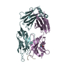
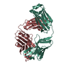
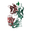
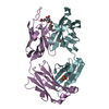
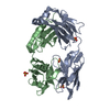
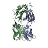
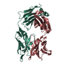
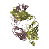
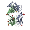
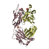
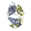
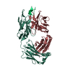
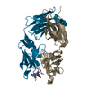
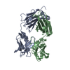
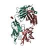
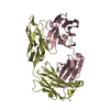
| Entry | Database: PDB / ID: 1bln | ||||||
|---|---|---|---|---|---|---|---|
| Title | ANTI-P-GLYCOPROTEIN FAB MRK-16 | ||||||
 Components Components |
| ||||||
 Keywords Keywords | IMMUNE SYSTEM / IMMUNOGLOBULIN | ||||||
| Function / homology | Immunoglobulins / Immunoglobulin-like / Sandwich / Mainly Beta Function and homology information Function and homology information | ||||||
| Biological species |  | ||||||
| Method |  X-RAY DIFFRACTION / MOLECULAR REPLACEMENT / Resolution: 2.8 Å X-RAY DIFFRACTION / MOLECULAR REPLACEMENT / Resolution: 2.8 Å | ||||||
 Authors Authors | Vasudevan, S. / Tsuruo, T. / Rose, D.R. | ||||||
 Citation Citation | Journal: J.Biol.Chem. / Year: 1998 Title: Mode of binding of anti-P-glycoprotein antibody MRK-16 to its antigen. A crystallographic and molecular modeling study. Authors: Vasudevan, S. / Tsuruo, T. / Rose, D.R. #1: Journal: Protein Pept.Lett. / Year: 1996Title: Preliminary Crystallographic Analysis of Anti-P-Gl Fab MRK-16 in Complex with its Antigenic Peptide Authors: Vasudevan, S. / Johns, K. / Rose, D.R. | ||||||
| History |
|
- Structure visualization
| Structure viewer | Molecule: MolmilJmol/JSmol |
|---|
- Downloads & links
-Download
| PDBx/mmCIF format | 1bln.cif.gz | 168.3 KB | Display | PDBx/mmCIF format |
|---|---|---|---|---|
| PDB format | pdb1bln.ent.gz | 133.9 KB | Display | PDB format |
| PDBx/mmJSON format | 1bln.json.gz | Tree view | PDBx/mmJSON format | |
| Others |  Other downloads Other downloads |
-Validation report
| Arichive directory | https://data.pdbj.org/pub/pdb/validation_reports/bl/1blnftp://data.pdbj.org/pub/pdb/validation_reports/bl/1bln | HTTPS FTP |
|---|
-Related structure data
| Related structure data |  1mcpS S: Starting model for refinement |
|---|---|
| Similar structure data |


-Links
 PDBj
PDBj


- Assembly
| Deposited unit | 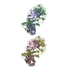
| ||||||||
|---|---|---|---|---|---|---|---|---|---|
| 1 | 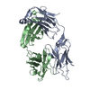
| ||||||||
| 2 | 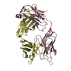
| ||||||||
| Unit cell |
| ||||||||
| Noncrystallographic symmetry (NCS) | NCS oper: (Code: given Matrix: (0.9883, 0.1522, 0.0131), Vector: |
-Components
| #1: Antibody | Mass: 23583.145 Da / Num. of mol.: 2 / Fragment: FAB / Source method: isolated from a natural source / Source: (natural) #2: Antibody | Mass: 23415.236 Da / Num. of mol.: 2 / Fragment: FAB / Source method: isolated from a natural source / Source: (natural) Has protein modification | Y | |
|---|
-Experimental details
-Experiment
| Experiment | Method: X-RAY DIFFRACTION / Number of used crystals: 1 |
|---|
- Sample preparation
| Crystal | Density Matthews: 2.14 Å3/Da / Density % sol: 53 % |
|---|---|
| Crystal grow | pH: 4 / Details: pH 4.0 |
| Crystal grow | *PLUS Method: other / Details: Vasudevan, S., (1994) J. Mol. Biol., 241, 736. |
-Data collection
| Diffraction | Mean temperature: 293 K |
|---|---|
| Diffraction source | Source: ROTATING ANODE / Type: RIGAKU RU200 / Wavelength: 1.5418 |
| Detector | Type: SDMS / Detector: AREA DETECTOR |
| Radiation | Monochromator: QUARTZ / Protocol: SINGLE WAVELENGTH / Monochromatic (M) / Laue (L): M / Scattering type: x-ray |
| Radiation wavelength | Wavelength: 1.5418 Å / Relative weight: 1 |
| Reflection | Resolution: 2.8→20 Å / Num. obs: 18167 / % possible obs: 86 % / Observed criterion σ(I): 1 / Redundancy: 4 % / Rsym value: 0.114 / Net I/σ(I): 9.7 |
| Reflection shell | Resolution: 2.8→3 Å / Redundancy: 1.6 % / Mean I/σ(I) obs: 2.1 / Rsym value: 0.22 / % possible all: 75 |
| Reflection | *PLUS Lowest resolution: 20 Å / % possible obs: 86 % / Num. measured all: 56513 / Rmerge(I) obs: 0.114 |
| Reflection shell | *PLUS % possible obs: 75 % / Num. unique obs: 3111 / Num. measured obs: 5374 / Rmerge(I) obs: 0.22 |
- Processing
| Software |
| ||||||||||||||||||||||||||||||||||||||||||||||||||||||||||||
|---|---|---|---|---|---|---|---|---|---|---|---|---|---|---|---|---|---|---|---|---|---|---|---|---|---|---|---|---|---|---|---|---|---|---|---|---|---|---|---|---|---|---|---|---|---|---|---|---|---|---|---|---|---|---|---|---|---|---|---|---|---|
| Refinement | Method to determine structure: MOLECULAR REPLACEMENT Starting model: 1MCP Resolution: 2.8→8 Å / Cross valid method: THROUGHOUT / σ(F): 1
| ||||||||||||||||||||||||||||||||||||||||||||||||||||||||||||
| Displacement parameters | Biso mean: 19 Å2 | ||||||||||||||||||||||||||||||||||||||||||||||||||||||||||||
| Refinement step | Cycle: LAST / Resolution: 2.8→8 Å
| ||||||||||||||||||||||||||||||||||||||||||||||||||||||||||||
| Refine LS restraints |
| ||||||||||||||||||||||||||||||||||||||||||||||||||||||||||||
| Xplor file | Serial no: 1 / Param file: PARAM19X.PRO / Topol file: TOPH19X.PRO | ||||||||||||||||||||||||||||||||||||||||||||||||||||||||||||
| Refinement | *PLUS % reflection Rfree: 5 % | ||||||||||||||||||||||||||||||||||||||||||||||||||||||||||||
| Solvent computation | *PLUS | ||||||||||||||||||||||||||||||||||||||||||||||||||||||||||||
| Displacement parameters | *PLUS Biso mean: 19 Å2 | ||||||||||||||||||||||||||||||||||||||||||||||||||||||||||||
| Refine LS restraints | *PLUS
|
