type II site-specific deoxyribonuclease / type II site-specific deoxyribonuclease activity / metal ion binding / DNA / DNA (> 10) / Restriction endonuclease PfoI
Function and homology information
Biological species
Pseudomonas fluorescens (bacteria)
Method
X-RAY DIFFRACTION / SYNCHROTRON / MAD / Resolution: 2.284 Å
Evidence: gel filtration, The dimeric assembly is evidenced by gel filtration, dynamic light scattering, crystal structure and from the homology to other related restriction endonucleases
Type
Name
Symmetry operation
Number
identity operation
1_555
x,y,z
1
Buried area
13620 Å2
ΔGint
-133 kcal/mol
Surface area
25820 Å2
Method
PISA
Unit cell
Length a, b, c (Å)
56.279, 91.663, 152.742
Angle α, β, γ (deg.)
90.00, 90.00, 90.00
Int Tables number
19
Space group name H-M
P212121
-
Components
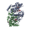
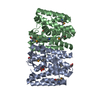
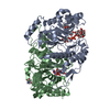
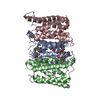
#1: Protein
RestrictionendonucleasePfoI
Mass: 35587.695 Da / Num. of mol.: 2 / Mutation: K187A Source method: isolated from a genetically manipulated source Source: (gene. exp.) Pseudomonas fluorescens (bacteria) / Variant: biovar 126 / Plasmid: pBAD24 / Production host: Escherichia coli (E. coli) / Strain (production host): ER2566 References: UniProt: A0A452CST9*PLUS, type II site-specific deoxyribonuclease
#2: DNA chain
DNA (5'-D(*CP*GP*CP*TP*CP*CP*CP*GP*GP*AP*GP*CP*GP*T)-3')
Mass: 4257.754 Da / Num. of mol.: 2 / Source method: obtained synthetically Details: Cognate oligoduplex with 3'-T-overhang and unpaired central recognition sequence base pair (C-C) Source: (synth.) Pseudomonas fluorescens (bacteria)
In the structure databanks used in Yorodumi, some data are registered as the other names, "COVID-19 virus" and "2019-nCoV". Here are the details of the virus and the list of structure data.
Jan 31, 2019. EMDB accession codes are about to change! (news from PDBe EMDB page)
EMDB accession codes are about to change! (news from PDBe EMDB page)
The allocation of 4 digits for EMDB accession codes will soon come to an end. Whilst these codes will remain in use, new EMDB accession codes will include an additional digit and will expand incrementally as the available range of codes is exhausted. The current 4-digit format prefixed with “EMD-” (i.e. EMD-XXXX) will advance to a 5-digit format (i.e. EMD-XXXXX), and so on. It is currently estimated that the 4-digit codes will be depleted around Spring 2019, at which point the 5-digit format will come into force.
The EM Navigator/Yorodumi systems omit the EMD- prefix.
Related info.:Q: What is EMD? / ID/Accession-code notation in Yorodumi/EM Navigator
Yorodumi is a browser for structure data from EMDB, PDB, SASBDB, etc.
This page is also the successor to EM Navigator detail page, and also detail information page/front-end page for Omokage search.
The word "yorodu" (or yorozu) is an old Japanese word meaning "ten thousand". "mi" (miru) is to see.
Related info.:EMDB / PDB / SASBDB / Comparison of 3 databanks / Yorodumi Search / Aug 31, 2016. New EM Navigator & Yorodumi / Yorodumi Papers / Jmol/JSmol / Function and homology information / Changes in new EM Navigator and Yorodumi
 Movie
Movie Controller

 Yorodumi
 Open data
 Basic information
 Components
 Keywords
 Function and homology information
 Pseudomonas fluorescens (bacteria)
 X-RAY DIFFRACTION /
  Authors
 Citation
 Structure visualization
 Downloads & links
 Other downloads


 PDBj
PDBj


 Assembly

 Mass: 40.078 Da / Num. of mol.: 4 / Source method: obtained synthetically / Formula: Ca
 Mass: 18.015 Da / Num. of mol.: 263 / Source method: isolated from a natural source / Formula: H2O
 Sample preparation
 / Beamline: I911-3 / Wavelength: 0.96112,0.98010
 Processing