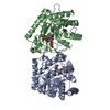
 Movie
Movie Controller


+ Open data

- Basic information
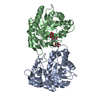
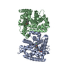
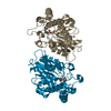
| Entry | Database: PDB / ID: 6ek1 | ||||||
|---|---|---|---|---|---|---|---|
| Title | Crystal structure of Type IIP restriction endonuclease PfoI | ||||||
 Components Components | restriction endonuclease PfoI | ||||||
 Keywords Keywords | HYDROLASE / Restriction endonuclease / PD-(D/E)xK nuclease | ||||||
| Function / homology | type II site-specific deoxyribonuclease / type II site-specific deoxyribonuclease activity / Restriction endonuclease PfoI Function and homology information Function and homology information | ||||||
| Biological species |  Pseudomonas fluorescens (bacteria) Pseudomonas fluorescens (bacteria) | ||||||
| Method |  X-RAY DIFFRACTION / MOLECULAR REPLACEMENT / Resolution: 2.601 Å X-RAY DIFFRACTION / MOLECULAR REPLACEMENT / Resolution: 2.601 Å | ||||||
 Authors Authors | Tamulaitiene, G. / Manakova, E. / Jovaisaite, V. / Grazulis, S. / Siksnys, V. | ||||||
| Funding support | Lithuania, 1items
| ||||||
 Citation Citation | Journal: Nucleic Acids Res. / Year: 2019 Title: Unique mechanism of target recognition by PfoI restriction endonuclease of the CCGG-family. Authors: Tamulaitiene, G. / Manakova, E. / Jovaisaite, V. / Tamulaitis, G. / Grazulis, S. / Bochtler, M. / Siksnys, V. | ||||||
| History |
|
- Structure visualization
| Structure viewer | Molecule: MolmilJmol/JSmol |
|---|
- Downloads & links
-Download
| PDBx/mmCIF format | 6ek1.cif.gz | 72.7 KB | Display | PDBx/mmCIF format |
|---|---|---|---|---|
| PDB format | pdb6ek1.ent.gz | 53.6 KB | Display | PDB format |
| PDBx/mmJSON format | 6ek1.json.gz | Tree view | PDBx/mmJSON format | |
| Others |  Other downloads Other downloads |
-Validation report
| Arichive directory | https://data.pdbj.org/pub/pdb/validation_reports/ek/6ek1ftp://data.pdbj.org/pub/pdb/validation_reports/ek/6ek1 | HTTPS FTP |
|---|
-Related structure data


-Links
 PDBj
PDBj

- Assembly
| Deposited unit | 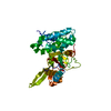
| ||||||||
|---|---|---|---|---|---|---|---|---|---|
| 1 | 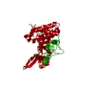
| ||||||||
| Unit cell |
|
-Components
| #1: Protein | Mass: 35194.527 Da / Num. of mol.: 1 Source method: isolated from a genetically manipulated source Source: (gene. exp.) Pseudomonas fluorescens (bacteria) / Strain: biovar 126 / Plasmid: pBAD24 / Production host: References: UniProt: A0A452CST7*PLUS, type II site-specific deoxyribonuclease |
|---|---|
| #2: Chemical | ChemComp-EDO /   Mass: 62.068 Da / Num. of mol.: 1 / Source method: obtained synthetically / Formula: C2H6O2 Mass: 62.068 Da / Num. of mol.: 1 / Source method: obtained synthetically / Formula: C2H6O2 |
| #3: Water | ChemComp-HOH /  Mass: 18.015 Da / Num. of mol.: 6 / Source method: isolated from a natural source / Formula: H2O Mass: 18.015 Da / Num. of mol.: 6 / Source method: isolated from a natural source / Formula: H2O |
-Experimental details
-Experiment
| Experiment | Method: X-RAY DIFFRACTION / Number of used crystals: 1 |
|---|
- Sample preparation
| Crystal | Density Matthews: 2.19 Å3/Da / Density % sol: 43.84 % / Description: Bipyramidal crystals |
|---|---|
| Crystal grow | Temperature: 292 K / Method: vapor diffusion Details: 6.8 mg/ml of PfoI in complex with DNA oligoduplex was mixed with reservoir solution 1:1, microseeding was used. NaHepes 0.1M, magnesium formate 0.2M, 18-20% PEG3350 PH range: 7-7.5 |
-Data collection
| Diffraction | Mean temperature: 100 K |
|---|---|
| Diffraction source | Source: ROTATING ANODE / Type: RIGAKU RUH3R / Wavelength: 1.54 Å |
| Detector | Type: RIGAKU RAXIS IV++ / Detector: IMAGE PLATE / Date: May 9, 2009 / Details: OSMIC CONFOCAL MIRRORS |
| Radiation | Protocol: SINGLE WAVELENGTH / Monochromatic (M) / Laue (L): M / Scattering type: x-ray |
| Radiation wavelength | Wavelength: 1.54 Å / Relative weight: 1 |
| Reflection | Resolution: 2.6→45.22 Å / Num. obs: 17795 / % possible obs: 98.6 % / Redundancy: 5.3 % / Biso Wilson estimate: 49.262 Å2 / Rmerge(I) obs: 0.112 / Rpim(I) all: 0.074 / Rsym value: 0.112 / Net I/σ(I): 5.7 |
| Reflection shell | Resolution: 2.6→2.74 Å / Redundancy: 6.2 % / Rmerge(I) obs: 0.44 / Mean I/σ(I) obs: 3.4 / Num. unique obs: 1470 / Rsym value: 0.44 / % possible all: 100 |
- Processing
| Software |
| |||||||||||||||||||||||||||||||||||||||||||||||||||||||||||||||||||||||||||||||||||||||||||
|---|---|---|---|---|---|---|---|---|---|---|---|---|---|---|---|---|---|---|---|---|---|---|---|---|---|---|---|---|---|---|---|---|---|---|---|---|---|---|---|---|---|---|---|---|---|---|---|---|---|---|---|---|---|---|---|---|---|---|---|---|---|---|---|---|---|---|---|---|---|---|---|---|---|---|---|---|---|---|---|---|---|---|---|---|---|---|---|---|---|---|---|---|
| Refinement | Method to determine structure: MOLECULAR REPLACEMENT / Resolution: 2.601→45.219 Å / SU ML: 0.34 / Cross valid method: FREE R-VALUE / σ(F): 1.2 / Phase error: 27.41
| |||||||||||||||||||||||||||||||||||||||||||||||||||||||||||||||||||||||||||||||||||||||||||
| Solvent computation | Shrinkage radii: 0.9 Å / VDW probe radii: 1.11 Å | |||||||||||||||||||||||||||||||||||||||||||||||||||||||||||||||||||||||||||||||||||||||||||
| Refinement step | Cycle: LAST / Resolution: 2.601→45.219 Å
| |||||||||||||||||||||||||||||||||||||||||||||||||||||||||||||||||||||||||||||||||||||||||||
| Refine LS restraints |
| |||||||||||||||||||||||||||||||||||||||||||||||||||||||||||||||||||||||||||||||||||||||||||
| LS refinement shell |
|
