 Movie
Movie Controller

[English] 日本語
 Yorodumi
- PDB-3wux: Crystal structure of unsaturated glucuronyl hydrolase mutant D115... -
+ Open data

- Basic information
| Entry | Database: PDB / ID: 3wux | ||||||
|---|---|---|---|---|---|---|---|
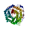
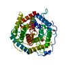
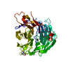
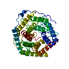
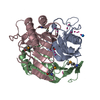
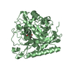
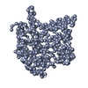
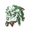
| Title | Crystal structure of unsaturated glucuronyl hydrolase mutant D115N/K370S from Streptococcus agalactiae | ||||||
 Components Components | Unsaturated chondroitin disaccharide hydrolase | ||||||
 Keywords Keywords | HYDROLASE / alpha6/alpha6-barrel | ||||||
| Function / homology |  Function and homology information Function and homology informationunsaturated chondroitin disaccharide hydrolase / unsaturated chondroitin disaccharide hydrolase activity / chondroitin hydrolase activity / polysaccharide catabolic process Similarity search - Function | ||||||
| Biological species |  Streptococcus agalactiae (bacteria) Streptococcus agalactiae (bacteria) | ||||||
| Method |  X-RAY DIFFRACTION / SYNCHROTRON / MOLECULAR REPLACEMENT / Resolution: 1.792 Å X-RAY DIFFRACTION / SYNCHROTRON / MOLECULAR REPLACEMENT / Resolution: 1.792 Å | ||||||
 Authors Authors | Nakamichi, Y. / Oiki, S. / Mikami, B. / Murata, K. / Hashimoto, W. | ||||||
 Citation Citation | Journal: to be published Title: Crystal structure of unsaturated glucuronyl hydrolase mutant D115N/K370S from Streptococcus agalactiae Authors: Nakamichi, Y. / Oiki, S. / Mikami, B. / Murata, K. / Hashimoto, W. | ||||||
| History |
|
- Structure visualization
| Structure viewer | Molecule: MolmilJmol/JSmol |
|---|
- Downloads & links
-Download
| PDBx/mmCIF format | 3wux.cif.gz | 95.2 KB | Display | PDBx/mmCIF format |
|---|---|---|---|---|
| PDB format | pdb3wux.ent.gz | 72.4 KB | Display | PDB format |
| PDBx/mmJSON format | 3wux.json.gz | Tree view | PDBx/mmJSON format | |
| Others |  Other downloads Other downloads |
-Validation report
| Arichive directory | https://data.pdbj.org/pub/pdb/validation_reports/wu/3wuxftp://data.pdbj.org/pub/pdb/validation_reports/wu/3wux | HTTPS FTP |
|---|
-Related structure data
| Related structure data |  3anjS S: Starting model for refinement |
|---|---|
| Similar structure data |


-Links
 PDBj
PDBj
- Assembly
| Deposited unit | 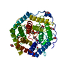
| ||||||||
|---|---|---|---|---|---|---|---|---|---|
| 1 |
| ||||||||
| 2 | 
| ||||||||
| Unit cell |
| ||||||||
| Components on special symmetry positions |
|
-Components
| #1: Protein | Mass: 46598.594 Da / Num. of mol.: 1 / Mutation: D115N/K370S Source method: isolated from a genetically manipulated source Source: (gene. exp.) Streptococcus agalactiae (bacteria) / Strain: NEM316 / Gene: gbs1889 / Plasmid: pET21b / Production host: References: UniProt: Q8E372, unsaturated chondroitin disaccharide hydrolase | ||
|---|---|---|---|
| #2: Chemical | ChemComp-EDO /   Mass: 62.068 Da / Num. of mol.: 12 / Source method: obtained synthetically / Formula: C2H6O2 Mass: 62.068 Da / Num. of mol.: 12 / Source method: obtained synthetically / Formula: C2H6O2#3: Water | ChemComp-HOH / |  Mass: 18.015 Da / Num. of mol.: 135 / Source method: isolated from a natural source / Formula: H2O Mass: 18.015 Da / Num. of mol.: 135 / Source method: isolated from a natural source / Formula: H2O |
-Experimental details
-Experiment
| Experiment | Method: X-RAY DIFFRACTION / Number of used crystals: 1 |
|---|
- Sample preparation
| Crystal | Density Matthews: 2.09 Å3/Da / Density % sol: 41.16 % |
|---|---|
| Crystal grow | Temperature: 293 K / Method: vapor diffusion, hanging drop / pH: 7 Details: 40% ethylene glycol, 0.1M tris(hydroxymethyl)aminomethane-HCl, 0.5mM dithiothreitol, pH 7.0, VAPOR DIFFUSION, HANGING DROP, temperature 293K |
-Data collection
| Diffraction | Mean temperature: 100 K |
|---|---|
| Diffraction source | Source: SYNCHROTRON / Site: SPring-8  / Beamline: BL38B1 / Wavelength: 1 Å / Beamline: BL38B1 / Wavelength: 1 Å |
| Detector | Type: ADSC QUANTUM 315r / Detector: CCD / Date: Dec 18, 2013 |
| Radiation | Monochromator: Si (111) double crystal / Protocol: SINGLE WAVELENGTH / Monochromatic (M) / Laue (L): M / Scattering type: x-ray |
| Radiation wavelength | Wavelength: 1 Å / Relative weight: 1 |
| Reflection | Resolution: 1.79→50 Å / Num. all: 38553 / Num. obs: 35858 / % possible obs: 98.5 % / Observed criterion σ(I): -3 / Biso Wilson estimate: 26.28 Å2 |
| Reflection shell | Resolution: 1.79→1.85 Å / % possible all: 94.9 |
- Processing
| Software |
| ||||||||||||||||||||||||||||||||||||||||||||||||||||||||||||||||||||||||||||||||||||||||||||||||||
|---|---|---|---|---|---|---|---|---|---|---|---|---|---|---|---|---|---|---|---|---|---|---|---|---|---|---|---|---|---|---|---|---|---|---|---|---|---|---|---|---|---|---|---|---|---|---|---|---|---|---|---|---|---|---|---|---|---|---|---|---|---|---|---|---|---|---|---|---|---|---|---|---|---|---|---|---|---|---|---|---|---|---|---|---|---|---|---|---|---|---|---|---|---|---|---|---|---|---|---|
| Refinement | Method to determine structure: MOLECULAR REPLACEMENT Starting model: 3ANJ Resolution: 1.792→29.056 Å / FOM work R set: 0.8164 / SU ML: 0.19 / σ(F): 1.33 / Phase error: 24.85 / Stereochemistry target values: ML
| ||||||||||||||||||||||||||||||||||||||||||||||||||||||||||||||||||||||||||||||||||||||||||||||||||
| Solvent computation | Shrinkage radii: 0.9 Å / VDW probe radii: 1.11 Å / Solvent model: FLAT BULK SOLVENT MODEL | ||||||||||||||||||||||||||||||||||||||||||||||||||||||||||||||||||||||||||||||||||||||||||||||||||
| Displacement parameters | Biso max: 80.53 Å2 / Biso mean: 29.31 Å2 / Biso min: 12.17 Å2 | ||||||||||||||||||||||||||||||||||||||||||||||||||||||||||||||||||||||||||||||||||||||||||||||||||
| Refinement step | Cycle: LAST / Resolution: 1.792→29.056 Å
| ||||||||||||||||||||||||||||||||||||||||||||||||||||||||||||||||||||||||||||||||||||||||||||||||||
| Refine LS restraints |
| ||||||||||||||||||||||||||||||||||||||||||||||||||||||||||||||||||||||||||||||||||||||||||||||||||
| LS refinement shell | Refine-ID: X-RAY DIFFRACTION / Total num. of bins used: 13
|
