 Movie
Movie Controller

[English] 日本語
 Yorodumi
- PDB-4z4j: Crystal Structure of cellobiose 2-epimerase from Caldicellulosiru... -
+ Open data

- Basic information
| Entry | Database: PDB / ID: 4z4j | ||||||
|---|---|---|---|---|---|---|---|
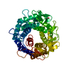
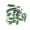
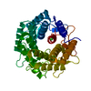
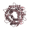
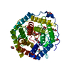
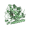
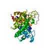
| Title | Crystal Structure of cellobiose 2-epimerase from Caldicellulosiruptor saccharolyticus DSM 8903 | ||||||
 Components Components | Cellobiose 2-epimerase | ||||||
 Keywords Keywords | ISOMERASE / Cellobiose 2-epimerase / latulose / lactose | ||||||
| Function / homology |  Function and homology information Function and homology informationcellobiose epimerase / cellobiose epimerase activity / carbohydrate metabolic process Similarity search - Function | ||||||
| Biological species |   Caldicellulosiruptor saccharolyticus DSM 8903 (bacteria) Caldicellulosiruptor saccharolyticus DSM 8903 (bacteria) | ||||||
| Method |  X-RAY DIFFRACTION / SYNCHROTRON / MIR / Resolution: 1.542 Å X-RAY DIFFRACTION / SYNCHROTRON / MIR / Resolution: 1.542 Å | ||||||
 Authors Authors | Shen, Q.Y. / Zhang, Y.Z. | ||||||
 Citation Citation | Journal: to be published Title: Crystal Structure of cellobiose 2-epimerase from Caldicellulosiruptor saccharolyticus DSM 8903 Authors: Shen, Q.Y. / Zhang, Y.Z. | ||||||
| History |
|
- Structure visualization
| Structure viewer | Molecule: MolmilJmol/JSmol |
|---|
- Downloads & links
-Download
| PDBx/mmCIF format | 4z4j.cif.gz | 191.6 KB | Display | PDBx/mmCIF format |
|---|---|---|---|---|
| PDB format | pdb4z4j.ent.gz | 151.4 KB | Display | PDB format |
| PDBx/mmJSON format | 4z4j.json.gz | Tree view | PDBx/mmJSON format | |
| Others |  Other downloads Other downloads |
-Validation report
| Arichive directory | https://data.pdbj.org/pub/pdb/validation_reports/z4/4z4jftp://data.pdbj.org/pub/pdb/validation_reports/z4/4z4j | HTTPS FTP |
|---|
-Related structure data


-Links
 PDBj
PDBj

- Assembly
| Deposited unit | 
| ||||||||
|---|---|---|---|---|---|---|---|---|---|
| 1 |
| ||||||||
| Unit cell |
|
-Components
| #1: Protein | Mass: 47764.355 Da / Num. of mol.: 1 Source method: isolated from a genetically manipulated source Source: (gene. exp.) Caldicellulosiruptor saccharolyticus DSM 8903 (bacteria)Strain: DSM8903 / Gene: Csac_0294 / Plasmid: pET28 / Production host: | ||||
|---|---|---|---|---|---|
| #2: Chemical |   Mass: 60.095 Da / Num. of mol.: 2 / Source method: obtained synthetically / Formula: C3H8O Mass: 60.095 Da / Num. of mol.: 2 / Source method: obtained synthetically / Formula: C3H8O#3: Chemical | ChemComp-EDO /   Mass: 62.068 Da / Num. of mol.: 4 / Source method: obtained synthetically / Formula: C2H6O2 Mass: 62.068 Da / Num. of mol.: 4 / Source method: obtained synthetically / Formula: C2H6O2#4: Water | ChemComp-HOH / |  Mass: 18.015 Da / Num. of mol.: 204 / Source method: isolated from a natural source / Formula: H2O Mass: 18.015 Da / Num. of mol.: 204 / Source method: isolated from a natural source / Formula: H2O |
-Experimental details
-Experiment
| Experiment | Method: X-RAY DIFFRACTION / Number of used crystals: 1 |
|---|
- Sample preparation
| Crystal | Density Matthews: 2.01 Å3/Da / Density % sol: 38.91 % |
|---|---|
| Crystal grow | Temperature: 293 K / Method: vapor diffusion, hanging drop / pH: 5.6 Details: 0.1 M Na citrate tribasic dihydrate pH 5.6, 20% v/v isopropanol, 20% v/v PEG 400 |
-Data collection
| Diffraction | Mean temperature: 110 K | |||||||||||||||||||||||||||||||||||||||||||||||||||||||||||||||||||||||||||||||||||||||||||||||||||||||||||||||||||||||||||||||||||||||||||||||||||||||||||||||||||||||||||||||||||||||||||||
|---|---|---|---|---|---|---|---|---|---|---|---|---|---|---|---|---|---|---|---|---|---|---|---|---|---|---|---|---|---|---|---|---|---|---|---|---|---|---|---|---|---|---|---|---|---|---|---|---|---|---|---|---|---|---|---|---|---|---|---|---|---|---|---|---|---|---|---|---|---|---|---|---|---|---|---|---|---|---|---|---|---|---|---|---|---|---|---|---|---|---|---|---|---|---|---|---|---|---|---|---|---|---|---|---|---|---|---|---|---|---|---|---|---|---|---|---|---|---|---|---|---|---|---|---|---|---|---|---|---|---|---|---|---|---|---|---|---|---|---|---|---|---|---|---|---|---|---|---|---|---|---|---|---|---|---|---|---|---|---|---|---|---|---|---|---|---|---|---|---|---|---|---|---|---|---|---|---|---|---|---|---|---|---|---|---|---|---|---|---|---|
| Diffraction source | Source: SYNCHROTRON / Site: APS  / Beamline: 23-ID-B / Wavelength: 0.968 Å / Beamline: 23-ID-B / Wavelength: 0.968 Å | |||||||||||||||||||||||||||||||||||||||||||||||||||||||||||||||||||||||||||||||||||||||||||||||||||||||||||||||||||||||||||||||||||||||||||||||||||||||||||||||||||||||||||||||||||||||||||||
| Detector | Type: MARMOSAIC 300 mm CCD / Detector: CCD / Date: Oct 25, 2014 | |||||||||||||||||||||||||||||||||||||||||||||||||||||||||||||||||||||||||||||||||||||||||||||||||||||||||||||||||||||||||||||||||||||||||||||||||||||||||||||||||||||||||||||||||||||||||||||
| Radiation | Monochromator: ID / Protocol: SINGLE WAVELENGTH / Monochromatic (M) / Laue (L): M / Scattering type: x-ray | |||||||||||||||||||||||||||||||||||||||||||||||||||||||||||||||||||||||||||||||||||||||||||||||||||||||||||||||||||||||||||||||||||||||||||||||||||||||||||||||||||||||||||||||||||||||||||||
| Radiation wavelength | Wavelength: 0.968 Å / Relative weight: 1 | |||||||||||||||||||||||||||||||||||||||||||||||||||||||||||||||||||||||||||||||||||||||||||||||||||||||||||||||||||||||||||||||||||||||||||||||||||||||||||||||||||||||||||||||||||||||||||||
| Reflection | Resolution: 1.54→30 Å / Num. obs: 55473 / % possible obs: 96.5 % / Redundancy: 13.8 % / Rmerge(I) obs: 0.1 / Rpim(I) all: 0.029 / Rrim(I) all: 0.104 / Χ2: 0.996 / Net I/av σ(I): 21.709 / Net I/σ(I): 12 / Num. measured all: 766957 | |||||||||||||||||||||||||||||||||||||||||||||||||||||||||||||||||||||||||||||||||||||||||||||||||||||||||||||||||||||||||||||||||||||||||||||||||||||||||||||||||||||||||||||||||||||||||||||
| Reflection shell | Diffraction-ID: 1 / Rejects: _
|
- Processing
| Software |
| ||||||||||||||||||||||||||||||||||||||||||||||||||||||||||||||||||||||||||||||||||||||||||||||||||||||||||||
|---|---|---|---|---|---|---|---|---|---|---|---|---|---|---|---|---|---|---|---|---|---|---|---|---|---|---|---|---|---|---|---|---|---|---|---|---|---|---|---|---|---|---|---|---|---|---|---|---|---|---|---|---|---|---|---|---|---|---|---|---|---|---|---|---|---|---|---|---|---|---|---|---|---|---|---|---|---|---|---|---|---|---|---|---|---|---|---|---|---|---|---|---|---|---|---|---|---|---|---|---|---|---|---|---|---|---|---|---|---|
| Refinement | Method to determine structure: MIR / Resolution: 1.542→29.625 Å / SU ML: 0.15 / Cross valid method: FREE R-VALUE / σ(F): 1.34 / Phase error: 17.6 / Stereochemistry target values: ML
| ||||||||||||||||||||||||||||||||||||||||||||||||||||||||||||||||||||||||||||||||||||||||||||||||||||||||||||
| Solvent computation | Shrinkage radii: 0.9 Å / VDW probe radii: 1.11 Å / Solvent model: FLAT BULK SOLVENT MODEL | ||||||||||||||||||||||||||||||||||||||||||||||||||||||||||||||||||||||||||||||||||||||||||||||||||||||||||||
| Displacement parameters | Biso max: 74.95 Å2 / Biso mean: 18.6559 Å2 / Biso min: 7.67 Å2 | ||||||||||||||||||||||||||||||||||||||||||||||||||||||||||||||||||||||||||||||||||||||||||||||||||||||||||||
| Refinement step | Cycle: final / Resolution: 1.542→29.625 Å
| ||||||||||||||||||||||||||||||||||||||||||||||||||||||||||||||||||||||||||||||||||||||||||||||||||||||||||||
| Refinement TLS params. | Method: refined / Origin x: 26.1514 Å / Origin y: 0.1351 Å / Origin z: -8.2895 Å
| ||||||||||||||||||||||||||||||||||||||||||||||||||||||||||||||||||||||||||||||||||||||||||||||||||||||||||||
| Refinement TLS group |
|
