 Movie
Movie Controller

[English] 日本語
 Yorodumi
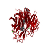
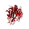
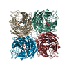
- PDB-1inf: INFLUENZA VIRUS B/LEE/40 NEURAMINIDASE COMPLEXED WITH BANA113 INH... -
+ Open data

- Basic information
| Entry | Database: PDB / ID: 1inf | |||||||||
|---|---|---|---|---|---|---|---|---|---|---|
| Title | INFLUENZA VIRUS B/LEE/40 NEURAMINIDASE COMPLEXED WITH BANA113 INHIBITOR | |||||||||
 Components Components | INFLUENZA VIRUS B/LEE/40 NEURAMINIDASE | |||||||||
 Keywords Keywords | HYDROLASE (O-GLYCOSYL) / NEURAMINIDASE / SIALIDASE / HYDROLASE / O-GLYCOSYL | |||||||||
| Function / homology |  Function and homology information Function and homology informationexo-alpha-sialidase / exo-alpha-sialidase activity / viral budding from plasma membrane / carbohydrate metabolic process / host cell plasma membrane / virion membrane / membrane / metal ion binding Similarity search - Function | |||||||||
| Biological species |  Influenza B virus Influenza B virus | |||||||||
| Method |  X-RAY DIFFRACTION / Resolution: 2.4 Å X-RAY DIFFRACTION / Resolution: 2.4 Å | |||||||||
 Authors Authors | Jedrzejas, M.J. / Luo, M. | |||||||||
 Citation Citation | Journal: J.Med.Chem. / Year: 1995 Title: Structure-based inhibitors of influenza virus sialidase. A benzoic acid lead with novel interaction. Authors: Singh, S. / Jedrzejas, M.J. / Air, G.M. / Luo, M. / Laver, W.G. / Brouillette, W.J. #1: Journal: Acta Crystallogr.,Sect.D / Year: 1995Title: Benzoic Acid Inhibitors of Influenza Virus Neuraminidase Authors: Luo, M. / Jedrzejas, M.J. / Singh, S. / White, C.L. / Brouillette, W.J. / Air, G.M. / Laver, W.G. #2: Journal: J.Med.Chem. / Year: 1995Title: Structure-Based Inhibitors of Influenza Viral Neuraminidase. A Benzoic Acid Lead with Novel Interaction Authors: Singh, S. / Jedrzejas, M.J. / Air, G.M. / Luo, M. / Laver, W.G. / Brouillette, W.J. #3: Journal: Biochemistry / Year: 1995Title: Structures of Aromatic Inhibitors of Influenza Virus Neuraminidase Authors: Jedrzejas, M.J. / Singh, S. / Brouillette, W.J. / Laver, W.G. / Air, G.M. / Luo, M. #4: Journal: Biochemistry / Year: 1994Title: Structure of Influenza Virus Neuraminidase B/Lee/40 Complexed with Sialic Acid and a Dehydro Analog at 1.8-A Resolution: Implications for the Catalytic Mechanism Authors: Janakiraman, M.N. / White, C.L. / Laver, W.G. / Air, G.M. / Luo, M. #5: Journal: J.Mol.Biol. / Year: 1991Title: Three-Dimensional Structure of the Neuraminidase of Influenza Virus A/Tokyo/3/67 at 2.2 A Resolution Authors: Varghese, J.N. / Colman, P.M. #6: Journal: J.Mol.Biol. / Year: 1991Title: Refined Atomic Structures of N9 Subtype Influenza Virus Neuraminidase and Escape Mutants Authors: Tulip, W.R. / Varghese, J.N. / Baker, A.T. / Van Donkelaar, A. / Laver, W.G. / Webster, R.G. / Colman, P.M. | |||||||||
| History |
|
- Structure visualization
| Structure viewer | Molecule: MolmilJmol/JSmol |
|---|
- Downloads & links
-Download
| PDBx/mmCIF format | 1inf.cif.gz | 113.9 KB | Display | PDBx/mmCIF format |
|---|---|---|---|---|
| PDB format | pdb1inf.ent.gz | 87.1 KB | Display | PDB format |
| PDBx/mmJSON format | 1inf.json.gz | Tree view | PDBx/mmJSON format | |
| Others |  Other downloads Other downloads |
-Validation report
| Arichive directory | https://data.pdbj.org/pub/pdb/validation_reports/in/1infftp://data.pdbj.org/pub/pdb/validation_reports/in/1inf | HTTPS FTP |
|---|
-Related structure data


-Links
 PDBj
PDBj

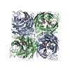
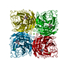
- Assembly
| Deposited unit | 
| ||||||||
|---|---|---|---|---|---|---|---|---|---|
| 1 | 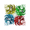
| ||||||||
| Unit cell |
| ||||||||
| Components on special symmetry positions |
|
-Components
| #1: Protein | Mass: 43432.332 Da / Num. of mol.: 1 / Source method: isolated from a natural source / Source: (natural) Influenza B virus / Genus: Influenzavirus B / Strain: STRAIN B-LEE-40 / References: UniProt: P03474, exo-alpha-sialidase | ||||||
|---|---|---|---|---|---|---|---|
| #2: Sugar | ChemComp-NAG /   Type: D-saccharide, beta linking / Mass: 221.208 Da / Num. of mol.: 1 Type: D-saccharide, beta linking / Mass: 221.208 Da / Num. of mol.: 1Source method: isolated from a genetically manipulated source Formula: C8H15NO6 | ||||||
| #3: Chemical |   Mass: 40.078 Da / Num. of mol.: 2 / Source method: obtained synthetically / Formula: Ca Mass: 40.078 Da / Num. of mol.: 2 / Source method: obtained synthetically / Formula: Ca#4: Chemical | ChemComp-ST4 / |   Mass: 238.243 Da / Num. of mol.: 1 / Source method: obtained synthetically / Formula: C10H14N4O3 Mass: 238.243 Da / Num. of mol.: 1 / Source method: obtained synthetically / Formula: C10H14N4O3#5: Water | ChemComp-HOH / |  Mass: 18.015 Da / Num. of mol.: 124 / Source method: isolated from a natural source / Formula: H2O Mass: 18.015 Da / Num. of mol.: 124 / Source method: isolated from a natural source / Formula: H2OHas protein modification | Y | |
-Experimental details
-Experiment
| Experiment | Method: X-RAY DIFFRACTION |
|---|
- Sample preparation
| Crystal | Density Matthews: 3.17 Å3/Da / Density % sol: 56 % | |||||||||||||||||||||||||||||||||||||||||||||||||
|---|---|---|---|---|---|---|---|---|---|---|---|---|---|---|---|---|---|---|---|---|---|---|---|---|---|---|---|---|---|---|---|---|---|---|---|---|---|---|---|---|---|---|---|---|---|---|---|---|---|---|
| Crystal grow | pH: 7.4 / Details: pH 7.4 | |||||||||||||||||||||||||||||||||||||||||||||||||
| Crystal | *PLUS Density % sol: 56 % | |||||||||||||||||||||||||||||||||||||||||||||||||
| Crystal grow | *PLUS Method: vapor diffusion, hanging drop / Details: Lin, Y., (1990) J. Mol. Biol., 214, 639. | |||||||||||||||||||||||||||||||||||||||||||||||||
| Components of the solutions | *PLUS
|
-Data collection
| Diffraction source | Wavelength: 1.5418 |
|---|---|
| Detector | Type: SIEMENS / Detector: AREA DETECTOR / Date: Apr 22, 1994 |
| Radiation | Monochromatic (M) / Laue (L): M / Scattering type: x-ray |
| Radiation wavelength | Wavelength: 1.5418 Å / Relative weight: 1 |
| Reflection | Num. obs: 17683 / Observed criterion σ(I): 1 / Redundancy: 4 % / Rmerge(I) obs: 0.125 |
| Reflection | *PLUS Highest resolution: 1.9 Å |
- Processing
| Software |
| ||||||||||||||||||||||||||||||||||||||||||||||||||||||||||||
|---|---|---|---|---|---|---|---|---|---|---|---|---|---|---|---|---|---|---|---|---|---|---|---|---|---|---|---|---|---|---|---|---|---|---|---|---|---|---|---|---|---|---|---|---|---|---|---|---|---|---|---|---|---|---|---|---|---|---|---|---|---|
| Refinement | Resolution: 2.4→6.5 Å / σ(F): 4 Details: THE REFINED MODEL COORDINATES DEPOSITED CONTAIN THE FULL PROTEIN SEQUENCE FROM GLU 77 TO LEU 466. THE TWO CALCIUM ATOMS ARE INCLUDED IN THE REFINED STRUCTURE. RESIDUE CA 500 STABILIZES A ...Details: THE REFINED MODEL COORDINATES DEPOSITED CONTAIN THE FULL PROTEIN SEQUENCE FROM GLU 77 TO LEU 466. THE TWO CALCIUM ATOMS ARE INCLUDED IN THE REFINED STRUCTURE. RESIDUE CA 500 STABILIZES A LOOP NEAR THE NEURAMINIDASE ACTIVE SITE, WHILE CA 501 IS LOCATED ON THE CRYSTALLOGRAPHIC NEURAMINIDASE TETRAMER FOUR-FOLD AXIS. THE EQUATORIAL PHOSPHONATE INHIBITOR IS RESIDUE EQP 500.
| ||||||||||||||||||||||||||||||||||||||||||||||||||||||||||||
| Refinement step | Cycle: LAST / Resolution: 2.4→6.5 Å
| ||||||||||||||||||||||||||||||||||||||||||||||||||||||||||||
| Refine LS restraints |
|
