 Movie
Movie Controller

[English] 日本語
 Yorodumi
- PDB-1b9v: NOVEL AROMATIC INHIBITORS OF INFLUENZA VIRUS NEURAMINIDASE MAKE S... -
+ Open data

- Basic information
| Entry | Database: PDB / ID: 1b9v | ||||||
|---|---|---|---|---|---|---|---|
| Title | NOVEL AROMATIC INHIBITORS OF INFLUENZA VIRUS NEURAMINIDASE MAKE SELECTIVE INTERACTIONS WITH CONSERVED RESIDUES AND WATER MOLECULES IN TEH ACTIVE SITE | ||||||
 Components Components | PROTEIN (NEURAMINIDASE) | ||||||
 Keywords Keywords | HYDROLASE / INFLUENZA / NEURAMINIDASE / SIALIDASE / B/LEE/40 | ||||||
| Function / homology |  Function and homology information Function and homology informationexo-alpha-sialidase / exo-alpha-sialidase activity / viral budding from plasma membrane / carbohydrate metabolic process / host cell plasma membrane / virion membrane / membrane / metal ion binding Similarity search - Function | ||||||
| Biological species |  Influenza B virus Influenza B virus | ||||||
| Method |  X-RAY DIFFRACTION / MOLECULAR REPLACEMENT / Resolution: 2.35 Å X-RAY DIFFRACTION / MOLECULAR REPLACEMENT / Resolution: 2.35 Å | ||||||
 Authors Authors | Finley, J.B. / Atigadda, V.R. / Duarte, F. / Zahao, J.J. / Brouillette, W.J. / Air, G.M. / Luo, M. | ||||||
 Citation Citation | Journal: J.Mol.Biol. / Year: 1999 Title: Novel aromatic inhibitors of influenza virus neuraminidase make selective interactions with conserved residues and water molecules in the active site. Authors: Finley, J.B. / Atigadda, V.R. / Duarte, F. / Zhao, J.J. / Brouillette, W.J. / Air, G.M. / Luo, M. | ||||||
| History |
|
- Structure visualization
| Structure viewer | Molecule: MolmilJmol/JSmol |
|---|
- Downloads & links
-Download
| PDBx/mmCIF format | 1b9v.cif.gz | 93 KB | Display | PDBx/mmCIF format |
|---|---|---|---|---|
| PDB format | pdb1b9v.ent.gz | 69.3 KB | Display | PDB format |
| PDBx/mmJSON format | 1b9v.json.gz | Tree view | PDBx/mmJSON format | |
| Others |  Other downloads Other downloads |
-Validation report
| Arichive directory | https://data.pdbj.org/pub/pdb/validation_reports/b9/1b9vftp://data.pdbj.org/pub/pdb/validation_reports/b9/1b9v | HTTPS FTP |
|---|
-Related structure data
| Related structure data |  1b9sC  1b9tC  1ivbS S: Starting model for refinement C: citing same article ( |
|---|---|
| Similar structure data |


-Links
 PDBj
PDBj

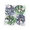
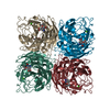
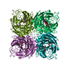
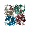
- Assembly
| Deposited unit | 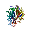
| ||||||||
|---|---|---|---|---|---|---|---|---|---|
| 1 | 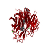
| ||||||||
| Unit cell |
|
-Components
| #1: Protein | Mass: 43460.348 Da / Num. of mol.: 1 / Source method: isolated from a natural source / Source: (natural) Influenza B virus (B/Lee/40) / Genus: Influenzavirus B / Species: Influenza B virus / Strain: B/Lee/40 / References: UniProt: P03474, exo-alpha-sialidase | ||||||
|---|---|---|---|---|---|---|---|
| #2: Sugar | ChemComp-NAG /   Type: D-saccharide, beta linking / Mass: 221.208 Da / Num. of mol.: 1 Type: D-saccharide, beta linking / Mass: 221.208 Da / Num. of mol.: 1Source method: isolated from a genetically manipulated source Formula: C8H15NO6 | ||||||
| #3: Chemical |   Mass: 40.078 Da / Num. of mol.: 2 / Source method: obtained synthetically / Formula: Ca Mass: 40.078 Da / Num. of mol.: 2 / Source method: obtained synthetically / Formula: Ca#4: Chemical | ChemComp-RA2 / |   Mass: 350.409 Da / Num. of mol.: 1 / Source method: obtained synthetically / Formula: C18H26N2O5 Mass: 350.409 Da / Num. of mol.: 1 / Source method: obtained synthetically / Formula: C18H26N2O5#5: Water | ChemComp-HOH / |  Mass: 18.015 Da / Num. of mol.: 107 / Source method: isolated from a natural source / Formula: H2O Mass: 18.015 Da / Num. of mol.: 107 / Source method: isolated from a natural source / Formula: H2OHas protein modification | Y | |
-Experimental details
-Experiment
| Experiment | Method: X-RAY DIFFRACTION / Number of used crystals: 1 |
|---|
- Sample preparation
| Crystal | Density Matthews: 3.2 Å3/Da / Density % sol: 61.56 % | |||||||||||||||||||||||||||||||||||||||||||||||||
|---|---|---|---|---|---|---|---|---|---|---|---|---|---|---|---|---|---|---|---|---|---|---|---|---|---|---|---|---|---|---|---|---|---|---|---|---|---|---|---|---|---|---|---|---|---|---|---|---|---|---|
| Crystal grow | pH: 6.8 / Details: pH 6.8 | |||||||||||||||||||||||||||||||||||||||||||||||||
| Crystal grow | *PLUS Method: vapor diffusion, hanging drop / Details: Sudbeck, E.A., (1997) J. Mol. Biol., 267, 584. | |||||||||||||||||||||||||||||||||||||||||||||||||
| Components of the solutions | *PLUS
|
-Data collection
| Diffraction | Mean temperature: 293 K |
|---|---|
| Diffraction source | Source: ROTATING ANODE / Type: RIGAKU RU200 / Wavelength: 1.5418 |
| Detector | Type: SIEMENS / Detector: AREA DETECTOR / Date: Aug 1, 1997 |
| Radiation | Monochromator: NI FILTER / Protocol: SINGLE WAVELENGTH / Monochromatic (M) / Laue (L): M / Scattering type: x-ray |
| Radiation wavelength | Wavelength: 1.5418 Å / Relative weight: 1 |
| Reflection | Resolution: 2.364→20 Å / Num. obs: 13395 / % possible obs: 80.15 % / Observed criterion σ(I): 3 / Redundancy: 2.32 % / Rsym value: 0.082 / Net I/σ(I): 9.98 |
| Reflection shell | Resolution: 2.236→2.448 Å / Redundancy: 2.32 % / Mean I/σ(I) obs: 3.17 / Rsym value: 0.228 / % possible all: 80.15 |
| Reflection | *PLUS Num. obs: 19008 / Rmerge(I) obs: 0.082 |
| Reflection shell | *PLUS % possible obs: 73.5 % / Rmerge(I) obs: 0.0023 |
- Processing
| Software |
| ||||||||||||||||||||||||||||||||||||||||||||||||||||||||||||
|---|---|---|---|---|---|---|---|---|---|---|---|---|---|---|---|---|---|---|---|---|---|---|---|---|---|---|---|---|---|---|---|---|---|---|---|---|---|---|---|---|---|---|---|---|---|---|---|---|---|---|---|---|---|---|---|---|---|---|---|---|---|
| Refinement | Method to determine structure: MOLECULAR REPLACEMENT Starting model: 1IVB Resolution: 2.35→8 Å / Cross valid method: THROUGHOUT / σ(F): 2
| ||||||||||||||||||||||||||||||||||||||||||||||||||||||||||||
| Refinement step | Cycle: LAST / Resolution: 2.35→8 Å
| ||||||||||||||||||||||||||||||||||||||||||||||||||||||||||||
| Refine LS restraints |
| ||||||||||||||||||||||||||||||||||||||||||||||||||||||||||||
| Software | *PLUS Name: X-PLOR / Version: 3.85 / Classification: refinement | ||||||||||||||||||||||||||||||||||||||||||||||||||||||||||||
| Refinement | *PLUS Lowest resolution: 8 Å / σ(F): 2 / Rfactor obs: 0.183 | ||||||||||||||||||||||||||||||||||||||||||||||||||||||||||||
| Solvent computation | *PLUS | ||||||||||||||||||||||||||||||||||||||||||||||||||||||||||||
| Displacement parameters | *PLUS | ||||||||||||||||||||||||||||||||||||||||||||||||||||||||||||
| LS refinement shell | *PLUS Rfactor Rfree: 0.577 / Rfactor obs: 0.364 |
