| Software | | Name | Version | Classification |
|---|
| Aimless | 0.3.11data scaling| PHENIX | 1.9_1692refinement| PDB_EXTRACT | 3.15 | data extraction| XDS | | data reduction| PHASER | | phasing | | | | | | |
|
|---|
| Refinement | Method to determine structure:  MOLECULAR REPLACEMENT MOLECULAR REPLACEMENT
Starting model: 4Z4J
Resolution: 1.671→29.59 Å / SU ML: 0.17 / Cross valid method: FREE R-VALUE / σ(F): 1.35 / Phase error: 18.67 / Stereochemistry target values: ML
| Rfactor | Num. reflection | % reflection |
|---|
| Rfree | 0.1979 | 1980 | 5.25 % |
|---|
| Rwork | 0.1631 | 35761 | - |
|---|
| obs | 0.1649 | 37741 | 83.07 % |
|---|
|
|---|
| Solvent computation | Shrinkage radii: 0.9 Å / VDW probe radii: 1.11 Å / Solvent model: FLAT BULK SOLVENT MODEL |
|---|
| Displacement parameters | Biso max: 90.76 Å2 / Biso mean: 28.2086 Å2 / Biso min: 13.64 Å2 |
|---|
| Refinement step | Cycle: final / Resolution: 1.671→29.59 Å
| Protein | Nucleic acid | Ligand | Solvent | Total |
|---|
| Num. atoms | 3285 | 0 | 4 | 74 | 3363 |
|---|
| Biso mean | - | - | 47.76 | 29.7 | - |
|---|
| Num. residues | - | - | - | - | 392 |
|---|
|
|---|
| Refine LS restraints | | Refine-ID | Type | Dev ideal | Number |
|---|
| X-RAY DIFFRACTION | f_bond_d| 0.007 | 3451 | | X-RAY DIFFRACTION | f_angle_d| 1.051 | 4664 | | X-RAY DIFFRACTION | f_chiral_restr| 0.044 | 483 | | X-RAY DIFFRACTION | f_plane_restr| 0.005 | 590 | | X-RAY DIFFRACTION | f_dihedral_angle_d| 15.031 | 1299 | | | | | |
|
|---|
| LS refinement shell | | Resolution (Å) | Rfactor Rfree | Num. reflection Rfree | Rfactor Rwork | Num. reflection Rwork | Refine-ID | % reflection obs (%) |
|---|
| 1.6712-1.713 | 0.2821 | 99 | 0.2338 | 2635 | X-RAY DIFFRACTION | 86 | | 1.713-1.7593 | 0.2569 | 198 | 0.2225 | 2550 | X-RAY DIFFRACTION | 86 | | 1.7593-1.8111 | 0.326 | 99 | 0.2067 | 2670 | X-RAY DIFFRACTION | 87 | | 1.8111-1.8695 | 0.2448 | 198 | 0.1944 | 2594 | X-RAY DIFFRACTION | 86 | | 1.8695-1.9363 | 0.2169 | 99 | 0.1852 | 2640 | X-RAY DIFFRACTION | 86 | | 1.9363-2.0138 | 0.2095 | 198 | 0.1758 | 2567 | X-RAY DIFFRACTION | 86 | | 2.0138-2.1055 | 0.208 | 99 | 0.1639 | 2636 | X-RAY DIFFRACTION | 85 | | 2.1055-2.2164 | 0.211 | 198 | 0.1615 | 2509 | X-RAY DIFFRACTION | 84 | | 2.2164-2.3552 | 0.2023 | 99 | 0.1629 | 2598 | X-RAY DIFFRACTION | 83 | | 2.3552-2.537 | 0.1878 | 169 | 0.1661 | 2526 | X-RAY DIFFRACTION | 83 | | 2.537-2.7921 | 0.1892 | 128 | 0.1678 | 2515 | X-RAY DIFFRACTION | 82 | | 2.7921-3.1957 | 0.2161 | 99 | 0.1734 | 2519 | X-RAY DIFFRACTION | 80 | | 3.1957-4.0247 | 0.1598 | 198 | 0.151 | 2375 | X-RAY DIFFRACTION | 78 | | 4.0247-29.59 | 0.2092 | 99 | 0.1372 | 2427 | X-RAY DIFFRACTION | 73 |
|
|---|
| Refinement TLS params. | Method: refined / Origin x: 26.3724 Å / Origin y: 0.1618 Å / Origin z: -8.2913 Å
| 11 | 12 | 13 | 21 | 22 | 23 | 31 | 32 | 33 |
|---|
| T | 0.1497 Å2 | -0.0091 Å2 | 0.0036 Å2 | - | 0.1807 Å2 | -0.0001 Å2 | - | - | 0.142 Å2 |
|---|
| L | 1.192 °2 | 0.13 °2 | -0.0524 °2 | - | 1.5241 °2 | -0.0808 °2 | - | - | 1.1448 °2 |
|---|
| S | 0.012 Å ° | 0.0358 Å ° | -0.0918 Å ° | 0.0017 Å ° | -0.0266 Å ° | -0.0283 Å ° | 0.0852 Å ° | -0.0574 Å ° | 0.013 Å ° |
|---|
|
|---|
| Refinement TLS group | | ID | Refine-ID | Refine TLS-ID | Selection details | Auth asym-ID | Auth seq-ID |
|---|
| 1 | X-RAY DIFFRACTION | 1 | allA| -1 - 390 | | 2 | X-RAY DIFFRACTION | 1 | allC| 1 - 68 | | 3 | X-RAY DIFFRACTION | 1 | allC| 69 - 74 | | 4 | X-RAY DIFFRACTION | 1 | allC| 75 | | 5 | X-RAY DIFFRACTION | 1 | allC| 76 - 78 | | 6 | X-RAY DIFFRACTION | 1 | allB| 1 | | | | | | | | | | | | |
|
|---|
 Movie
Movie Controller

 Yorodumi
 Open data
 Basic information
 Components
 Keywords
 Function and homology information
 Caldicellulosiruptor saccharolyticus DSM 8903 (bacteria)
 Authors
 Citation
 Structure visualization
 Downloads & links
 Other downloads
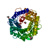


 PDBj
PDBj
 Assembly

 Mass: 62.068 Da / Num. of mol.: 1 / Source method: obtained synthetically / Formula: C2H6O2
 Mass: 18.015 Da / Num. of mol.: 74 / Source method: isolated from a natural source / Formula: H2O
 Sample preparation
 / Beamline: 23-ID-B / Wavelength: 0.968 Å
 Processing