 Movie
Movie Controller

[English] 日本語
 Yorodumi
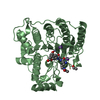
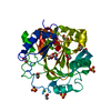
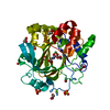
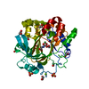
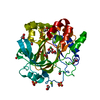
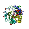
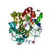
- PDB-1rh9: Family GH5 endo-beta-mannanase from Lycopersicon esculentum (tomato) -
+ Open data

- Basic information
| Entry | Database: PDB / ID: 1rh9 | ||||||
|---|---|---|---|---|---|---|---|
| Title | Family GH5 endo-beta-mannanase from Lycopersicon esculentum (tomato) | ||||||
 Components Components | endo-beta-mannanase | ||||||
 Keywords Keywords | HYDROLASE / ENDO-BETA-MANNASE / Retaining / Glycoside Hydrolase Family 5 / mannan | ||||||
| Function / homology |  Function and homology information Function and homology informationmannan endo-1,4-beta-mannosidase / mannan endo-1,4-beta-mannosidase activity / extracellular region Similarity search - Function | ||||||
| Biological species |  | ||||||
| Method |  X-RAY DIFFRACTION / SYNCHROTRON / MOLECULAR REPLACEMENT / Resolution: 1.5 Å X-RAY DIFFRACTION / SYNCHROTRON / MOLECULAR REPLACEMENT / Resolution: 1.5 Å | ||||||
 Authors Authors | Oakley, A.J. / Bourgault, R. / Bewley, J.D. / Wilce, M.C.J. | ||||||
 Citation Citation | Journal: Protein Sci. / Year: 2005 Title: Three-dimensional structure of (1,4)-beta-D-mannan mannanohydrolase from tomato fruit. Authors: Bourgault, R. / Oakley, A.J. / Bewley, J.D. / Wilce, M.C. | ||||||
| History |
|
- Structure visualization
| Structure viewer | Molecule: MolmilJmol/JSmol |
|---|
- Downloads & links
-Download
| PDBx/mmCIF format | 1rh9.cif.gz | 87.6 KB | Display | PDBx/mmCIF format |
|---|---|---|---|---|
| PDB format | pdb1rh9.ent.gz | 66.4 KB | Display | PDB format |
| PDBx/mmJSON format | 1rh9.json.gz | Tree view | PDBx/mmJSON format | |
| Others |  Other downloads Other downloads |
-Validation report
| Arichive directory | https://data.pdbj.org/pub/pdb/validation_reports/rh/1rh9ftp://data.pdbj.org/pub/pdb/validation_reports/rh/1rh9 | HTTPS FTP |
|---|
-Related structure data
| Related structure data |  1qnpS S: Starting model for refinement |
|---|---|
| Similar structure data |


-Links
 PDBj
PDBj- Assembly
| Deposited unit | 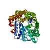
| ||||||||
|---|---|---|---|---|---|---|---|---|---|
| 1 |
| ||||||||
| Unit cell |
| ||||||||
| Details | The asymmetric unit contains the biological assembly (a monomer). |
-Components
| #1: Protein | Mass: 42435.910 Da / Num. of mol.: 1 / Fragment: residues 27-399 Source method: isolated from a genetically manipulated source Source: (gene. exp.)  References: UniProt: Q93WT4, UniProt: Q8L5J1*PLUS, mannan endo-1,4-beta-mannosidase |
|---|---|
| Has protein modification | Y |
-Experimental details
-Experiment
| Experiment | Method: X-RAY DIFFRACTION / Number of used crystals: 1 |
|---|
- Sample preparation
| Crystal | Density Matthews: 1.99 Å3/Da / Density % sol: 37.76 % |
|---|---|
| Crystal grow | Temperature: 298 K / Method: vapor diffusion, hanging drop / pH: 5.6 Details: 30% PEG 4000, 0.1M Na Citrate, 0.2M NH4 Acetate, pH 5.6, VAPOR DIFFUSION, HANGING DROP, temperature 298K |
-Data collection
| Diffraction | Mean temperature: 100 K |
|---|---|
| Diffraction source | Source: SYNCHROTRON / Site: APS  / Beamline: 14-BM-C / Wavelength: 0.9 Å / Beamline: 14-BM-C / Wavelength: 0.9 Å |
| Detector | Type: ADSC QUANTUM 4 / Detector: CCD / Date: Mar 1, 2003 / Details: BEAMLINE OPTICS |
| Radiation | Monochromator: BEAMLINE OPTICS / Protocol: SINGLE WAVELENGTH / Monochromatic (M) / Laue (L): M / Scattering type: x-ray |
| Radiation wavelength | Wavelength: 0.9 Å / Relative weight: 1 |
| Reflection | Resolution: 1.5→54.233 Å / Num. all: 56178 / Num. obs: 56178 / % possible obs: 98.4 % / Observed criterion σ(F): -1 / Observed criterion σ(I): -1 / Rmerge(I) obs: 0.08 / Net I/σ(I): 12.1 |
| Reflection shell | Resolution: 1.5→1.55 Å / Rmerge(I) obs: 0.743 / Mean I/σ(I) obs: 1.2 / % possible all: 99.7 |
- Processing
| Software |
| ||||||||||||||||||||||||||||||||||||||||||||||||||||||||||||||||||||||||||||||||||||||||||||||||||||
|---|---|---|---|---|---|---|---|---|---|---|---|---|---|---|---|---|---|---|---|---|---|---|---|---|---|---|---|---|---|---|---|---|---|---|---|---|---|---|---|---|---|---|---|---|---|---|---|---|---|---|---|---|---|---|---|---|---|---|---|---|---|---|---|---|---|---|---|---|---|---|---|---|---|---|---|---|---|---|---|---|---|---|---|---|---|---|---|---|---|---|---|---|---|---|---|---|---|---|---|---|---|
| Refinement | Method to determine structure: MOLECULAR REPLACEMENT Starting model: 1QNP Resolution: 1.5→20 Å / Cor.coef. Fo:Fc: 0.97 / Cor.coef. Fo:Fc free: 0.962 / SU B: 1.977 / SU ML: 0.068 / Cross valid method: THROUGHOUT / σ(F): -1 / ESU R: 0.084 / ESU R Free: 0.081 / Stereochemistry target values: MAXIMUM LIKELIHOOD / Details: HYDROGENS HAVE BEEN ADDED IN THE RIDING POSITIONS
| ||||||||||||||||||||||||||||||||||||||||||||||||||||||||||||||||||||||||||||||||||||||||||||||||||||
| Solvent computation | Ion probe radii: 0.8 Å / Shrinkage radii: 0.8 Å / VDW probe radii: 1.4 Å / Solvent model: BABINET MODEL WITH MASK | ||||||||||||||||||||||||||||||||||||||||||||||||||||||||||||||||||||||||||||||||||||||||||||||||||||
| Displacement parameters | Biso mean: 18.865 Å2
| ||||||||||||||||||||||||||||||||||||||||||||||||||||||||||||||||||||||||||||||||||||||||||||||||||||
| Refinement step | Cycle: LAST / Resolution: 1.5→20 Å
| ||||||||||||||||||||||||||||||||||||||||||||||||||||||||||||||||||||||||||||||||||||||||||||||||||||
| Refine LS restraints |
| ||||||||||||||||||||||||||||||||||||||||||||||||||||||||||||||||||||||||||||||||||||||||||||||||||||
| LS refinement shell | Resolution: 1.5→1.539 Å / Total num. of bins used: 20 /
|
