 Movie
Movie Controller


+ Open data

- Basic information
| Entry | Database: PDB / ID: 1bqc | ||||||
|---|---|---|---|---|---|---|---|
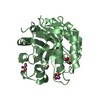
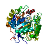
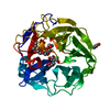
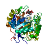
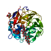
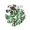
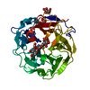
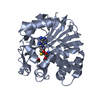
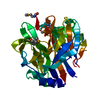
| Title | BETA-MANNANASE FROM THERMOMONOSPORA FUSCA | ||||||
 Components Components | PROTEIN (BETA-MANNANASE) | ||||||
 Keywords Keywords | HYDROLASE / MANNANASE / GLYCOSYL HYDROLASE / FAMILY 5 / THERMOMONOSPORA FUSCA | ||||||
| Function / homology |  Function and homology information Function and homology informationmannan endo-1,4-beta-mannosidase / mannan endo-1,4-beta-mannosidase activity / polysaccharide catabolic process Similarity search - Function | ||||||
| Biological species |   Thermobifida fusca (bacteria) Thermobifida fusca (bacteria) | ||||||
| Method |  X-RAY DIFFRACTION / SYNCHROTRON / MIRAS / Resolution: 1.5 Å X-RAY DIFFRACTION / SYNCHROTRON / MIRAS / Resolution: 1.5 Å | ||||||
 Authors Authors | Hilge, M. / Gloor, S.M. / Piontek, K. | ||||||
 Citation Citation | Journal: Structure / Year: 1998 Title: High-resolution native and complex structures of thermostable beta-mannanase from Thermomonospora fusca - substrate specificity in glycosyl hydrolase family 5. Authors: Hilge, M. / Gloor, S.M. / Rypniewski, W. / Sauer, O. / Heightman, T.D. / Zimmermann, W. / Winterhalter, K. / Piontek, K. #1: Journal: Acta Crystallogr.,Sect.D / Year: 2001 Title: Structure elucidation of beta-mannanase: from the electron-density map to the DNA sequence. Authors: Hilge, M. / Perrakis, A. / Abrahams, J.P. / Winterhalter, K. / Piontek, K. / Gloor, S.M. | ||||||
| History |
|
- Structure visualization
| Structure viewer | Molecule: MolmilJmol/JSmol |
|---|
- Downloads & links
-Download
| PDBx/mmCIF format | 1bqc.cif.gz | 79.4 KB | Display | PDBx/mmCIF format |
|---|---|---|---|---|
| PDB format | pdb1bqc.ent.gz | 58.9 KB | Display | PDB format |
| PDBx/mmJSON format | 1bqc.json.gz | Tree view | PDBx/mmJSON format | |
| Others |  Other downloads Other downloads |
-Validation report
| Arichive directory | https://data.pdbj.org/pub/pdb/validation_reports/bq/1bqcftp://data.pdbj.org/pub/pdb/validation_reports/bq/1bqc | HTTPS FTP |
|---|
-Related structure data


-Links
 PDBj
PDBj- Assembly
| Deposited unit | 
| ||||||||
|---|---|---|---|---|---|---|---|---|---|
| 1 |
| ||||||||
| Unit cell |
|
-Components
| #1: Protein | Mass: 33123.312 Da / Num. of mol.: 1 / Source method: isolated from a natural source / Details: GERMAN COLLECTION OF MICROORGANISMS (DSM) / Source: (natural) Thermobifida fusca (bacteria) / Strain: KW3References: UniProt: Q9ZF13, mannan endo-1,4-beta-mannosidase |
|---|---|
| #2: Water | ChemComp-HOH /  Mass: 18.015 Da / Num. of mol.: 434 / Source method: isolated from a natural source / Formula: H2O Mass: 18.015 Da / Num. of mol.: 434 / Source method: isolated from a natural source / Formula: H2O |
| Has protein modification | Y |
-Experimental details
-Experiment
| Experiment | Method: X-RAY DIFFRACTION / Number of used crystals: 1 |
|---|
- Sample preparation
| Crystal | Density Matthews: 2.01 Å3/Da / Density % sol: 38.88 % | ||||||||||||||||||
|---|---|---|---|---|---|---|---|---|---|---|---|---|---|---|---|---|---|---|---|
| Crystal grow | pH: 7 / Details: 1.85M AMMONIUM SULFATE, 0.1M HEPES, PH 7.0 | ||||||||||||||||||
| Crystal grow | *PLUS Method: vapor diffusion, hanging drop | ||||||||||||||||||
| Components of the solutions | *PLUS
|
-Data collection
| Diffraction | Mean temperature: 100 K |
|---|---|
| Diffraction source | Source: SYNCHROTRON / Site: EMBL/DESY, HAMBURG  / Beamline: X11 / Wavelength: 0.9072 / Beamline: X11 / Wavelength: 0.9072 |
| Detector | Type: MARRESEARCH / Detector: IMAGE PLATE / Date: Mar 15, 1997 |
| Radiation | Protocol: SINGLE WAVELENGTH / Monochromatic (M) / Laue (L): M / Scattering type: x-ray |
| Radiation wavelength | Wavelength: 0.9072 Å / Relative weight: 1 |
| Reflection | Resolution: 1.5→32 Å / Num. obs: 42667 / % possible obs: 97.2 % / Observed criterion σ(I): 0 / Redundancy: 3.22 % / Rmerge(I) obs: 0.062 / Rsym value: 0.062 / Net I/σ(I): 17.2 |
| Reflection shell | Resolution: 1.5→1.55 Å / Redundancy: 2.5 % / Rmerge(I) obs: 0.331 / Mean I/σ(I) obs: 2.9 / Rsym value: 0.331 / % possible all: 92.2 |
| Reflection shell | *PLUS % possible obs: 92.2 % |
- Processing
| Software |
| |||||||||||||||||||||||||||||||||||||||||||||||||||||||||||||||
|---|---|---|---|---|---|---|---|---|---|---|---|---|---|---|---|---|---|---|---|---|---|---|---|---|---|---|---|---|---|---|---|---|---|---|---|---|---|---|---|---|---|---|---|---|---|---|---|---|---|---|---|---|---|---|---|---|---|---|---|---|---|---|---|---|
| Refinement | Method to determine structure: MIRAS / Resolution: 1.5→32 Å / Cross valid method: THROUGHOUT / σ(F): 0 Details: INDIVIDUAL, ANISOTROPIC B-FACTOR REFINEMENT USING REFMAC 4.0.1
| |||||||||||||||||||||||||||||||||||||||||||||||||||||||||||||||
| Displacement parameters | Biso mean: 15.5 Å2 | |||||||||||||||||||||||||||||||||||||||||||||||||||||||||||||||
| Refinement step | Cycle: LAST / Resolution: 1.5→32 Å
| |||||||||||||||||||||||||||||||||||||||||||||||||||||||||||||||
| Refine LS restraints |
| |||||||||||||||||||||||||||||||||||||||||||||||||||||||||||||||
| Software | *PLUS Name: REFMAC / Classification: refinement | |||||||||||||||||||||||||||||||||||||||||||||||||||||||||||||||
| Refinement | *PLUS σ(F): 0 / % reflection Rfree: 5 % / Rfactor obs: 0.119 | |||||||||||||||||||||||||||||||||||||||||||||||||||||||||||||||
| Solvent computation | *PLUS | |||||||||||||||||||||||||||||||||||||||||||||||||||||||||||||||
| Displacement parameters | *PLUS Biso mean: 15.5 Å2 |
