 Movie
Movie Controller


+ Open data

- Basic information
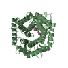
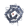
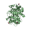
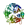
| Entry | Database: PDB / ID: 3vw5 | ||||||
|---|---|---|---|---|---|---|---|
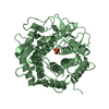
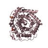
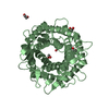
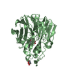
| Title | Crystal structure of sugar epimerase from ruminal bacterium | ||||||
 Components Components | Cellobiose 2-epimerase | ||||||
 Keywords Keywords | ISOMERASE / (alpha/alpha)6 barrel fold / Epimerase / Carbohydrate/Sugar Binding / Epimerization | ||||||
| Function / homology |  Function and homology information Function and homology informationcellobiose epimerase / cellobiose epimerase activity / carbohydrate metabolic process / cytoplasm Similarity search - Function | ||||||
| Biological species |  Ruminococcus albus (bacteria) Ruminococcus albus (bacteria) | ||||||
| Method |  X-RAY DIFFRACTION / SYNCHROTRON / MOLECULAR REPLACEMENT / Resolution: 2.6 Å X-RAY DIFFRACTION / SYNCHROTRON / MOLECULAR REPLACEMENT / Resolution: 2.6 Å | ||||||
 Authors Authors | Fujiwara, T. / Saburi, W. / Tanaka, I. / Yao, M. | ||||||
 Citation Citation | Journal: Febs Lett. / Year: 2013 Title: Crystal structure of Ruminococcus albus cellobiose 2-epimerase: structural insights into epimerization of unmodified sugar Authors: Fujiwara, T. / Saburi, W. / Inoue, S. / Mori, H. / Matsui, H. / Tanaka, I. / Yao, M. | ||||||
| History |
|
- Structure visualization
| Structure viewer | Molecule: MolmilJmol/JSmol |
|---|
- Downloads & links
-Download
| PDBx/mmCIF format | 3vw5.cif.gz | 241.1 KB | Display | PDBx/mmCIF format |
|---|---|---|---|---|
| PDB format | pdb3vw5.ent.gz | 195.9 KB | Display | PDB format |
| PDBx/mmJSON format | 3vw5.json.gz | Tree view | PDBx/mmJSON format | |
| Others |  Other downloads Other downloads |
-Validation report
| Arichive directory | https://data.pdbj.org/pub/pdb/validation_reports/vw/3vw5ftp://data.pdbj.org/pub/pdb/validation_reports/vw/3vw5 | HTTPS FTP |
|---|
-Related structure data
| Related structure data |  3gt5S S: Starting model for refinement |
|---|---|
| Similar structure data |


-Links
 PDBj
PDBj
- Assembly
| Deposited unit | 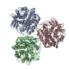
| ||||||||
|---|---|---|---|---|---|---|---|---|---|
| 1 | 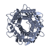
| ||||||||
| 2 | 
| ||||||||
| 3 | 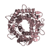
| ||||||||
| Unit cell |
|
-Components
| #1: Protein | Mass: 45362.727 Da / Num. of mol.: 3 / Mutation: G38E, P138L Source method: isolated from a genetically manipulated source Source: (gene. exp.) Ruminococcus albus (bacteria) / Strain: NE1 / Gene: CE / Plasmid: pET23A / Production host: References: UniProt: A8CF79, UniProt: P0DKY4*PLUS, cellobiose epimerase #2: Water | ChemComp-HOH / |  Mass: 18.015 Da / Num. of mol.: 156 / Source method: isolated from a natural source / Formula: H2O Mass: 18.015 Da / Num. of mol.: 156 / Source method: isolated from a natural source / Formula: H2O |
|---|
-Experimental details
-Experiment
| Experiment | Method: X-RAY DIFFRACTION / Number of used crystals: 1 |
|---|
- Sample preparation
| Crystal | Density Matthews: 3.41 Å3/Da / Density % sol: 63.93 % |
|---|---|
| Crystal grow | Temperature: 293 K / Method: vapor diffusion, sitting drop / pH: 7 Details: 2.6M sodium malonate, pH 7.0, VAPOR DIFFUSION, SITTING DROP, temperature 293K |
-Data collection
| Diffraction | Mean temperature: 100 K |
|---|---|
| Diffraction source | Source: SYNCHROTRON / Site: SPring-8  / Beamline: BL41XU / Wavelength: 1 Å / Beamline: BL41XU / Wavelength: 1 Å |
| Detector | Type: MARMOSAIC 225 mm CCD / Detector: CCD / Date: Jul 6, 2011 / Details: mirrors |
| Radiation | Monochromator: the rotated-inclined double-crystal monochromator Protocol: SINGLE WAVELENGTH / Monochromatic (M) / Laue (L): M / Scattering type: x-ray |
| Radiation wavelength | Wavelength: 1 Å / Relative weight: 1 |
| Reflection | Resolution: 2.6→45 Å / Num. obs: 57248 / % possible obs: 98.2 % / Redundancy: 3.7 % / Biso Wilson estimate: 48.981 Å2 / Rmerge(I) obs: 0.0085 / Net I/σ(I): 12.14 |
| Reflection shell | Resolution: 2.6→2.74 Å / Redundancy: 3.7 % / Rmerge(I) obs: 0.623 / Mean I/σ(I) obs: 2.1 / Num. unique all: 8838 / % possible all: 95.3 |
- Processing
| Software |
| ||||||||||||||||||||||||||||||||||||||||||||||||||||||||||||||||||||||||||||||||||||||||||||||||||||||||||||||||||||||||||||||||||||||||||||||||||||||||||
|---|---|---|---|---|---|---|---|---|---|---|---|---|---|---|---|---|---|---|---|---|---|---|---|---|---|---|---|---|---|---|---|---|---|---|---|---|---|---|---|---|---|---|---|---|---|---|---|---|---|---|---|---|---|---|---|---|---|---|---|---|---|---|---|---|---|---|---|---|---|---|---|---|---|---|---|---|---|---|---|---|---|---|---|---|---|---|---|---|---|---|---|---|---|---|---|---|---|---|---|---|---|---|---|---|---|---|---|---|---|---|---|---|---|---|---|---|---|---|---|---|---|---|---|---|---|---|---|---|---|---|---|---|---|---|---|---|---|---|---|---|---|---|---|---|---|---|---|---|---|---|---|---|---|---|---|
| Refinement | Method to determine structure: MOLECULAR REPLACEMENT Starting model: 3GT5 Resolution: 2.6→39.127 Å / SU ML: 0.63 / Isotropic thermal model: isotropic / Cross valid method: THROUGHOUT / Phase error: 21.82
| ||||||||||||||||||||||||||||||||||||||||||||||||||||||||||||||||||||||||||||||||||||||||||||||||||||||||||||||||||||||||||||||||||||||||||||||||||||||||||
| Solvent computation | Shrinkage radii: 0.83 Å / VDW probe radii: 1.1 Å / Solvent model: FLAT BULK SOLVENT MODEL / Bsol: 43.384 Å2 / ksol: 0.427 e/Å3 | ||||||||||||||||||||||||||||||||||||||||||||||||||||||||||||||||||||||||||||||||||||||||||||||||||||||||||||||||||||||||||||||||||||||||||||||||||||||||||
| Displacement parameters | Biso mean: 4.47 Å2
| ||||||||||||||||||||||||||||||||||||||||||||||||||||||||||||||||||||||||||||||||||||||||||||||||||||||||||||||||||||||||||||||||||||||||||||||||||||||||||
| Refinement step | Cycle: LAST / Resolution: 2.6→39.127 Å
| ||||||||||||||||||||||||||||||||||||||||||||||||||||||||||||||||||||||||||||||||||||||||||||||||||||||||||||||||||||||||||||||||||||||||||||||||||||||||||
| Refine LS restraints |
| ||||||||||||||||||||||||||||||||||||||||||||||||||||||||||||||||||||||||||||||||||||||||||||||||||||||||||||||||||||||||||||||||||||||||||||||||||||||||||
| LS refinement shell | Refine-ID: X-RAY DIFFRACTION / Total num. of bins used: 21
|
