 Movie
Movie Controller

[English] 日本語
 Yorodumi
- PDB-3vxd: Crystal structure of unsaturated glucuronyl hydrolase mutant D115... -
+ Open data

- Basic information
| Entry | Database: PDB / ID: 3vxd | ||||||
|---|---|---|---|---|---|---|---|
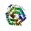
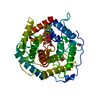
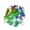
| Title | Crystal structure of unsaturated glucuronyl hydrolase mutant D115N from Streptcoccus agalactiae | ||||||
 Components Components | Putative uncharacterized protein gbs1889 | ||||||
 Keywords Keywords | HYDROLASE / alpha6/alpha6-barrel | ||||||
| Function / homology |  Function and homology information Function and homology informationunsaturated chondroitin disaccharide hydrolase / unsaturated chondroitin disaccharide hydrolase activity / chondroitin hydrolase activity / polysaccharide catabolic process Similarity search - Function | ||||||
| Biological species |  Streptococcus agalactiae (bacteria) Streptococcus agalactiae (bacteria) | ||||||
| Method |  X-RAY DIFFRACTION / SYNCHROTRON / MOLECULAR REPLACEMENT / Resolution: 2 Å X-RAY DIFFRACTION / SYNCHROTRON / MOLECULAR REPLACEMENT / Resolution: 2 Å | ||||||
 Authors Authors | Nakamichi, Y. / Maruyama, Y. / Mikami, B. / Hashimoto, W. / Murata, K. | ||||||
 Citation Citation | Journal: To be Published Title: Crystal structure of unsaturated glucuronyl hydrolase mutant D115N from Streptcoccus agalactiae Authors: Nakamichi, Y. / Maruyama, Y. / Mikami, B. / Hashimoto, W. / Murata, K. | ||||||
| History |
|
- Structure visualization
| Structure viewer | Molecule: MolmilJmol/JSmol |
|---|
- Downloads & links
-Download
| PDBx/mmCIF format | 3vxd.cif.gz | 315.6 KB | Display | PDBx/mmCIF format |
|---|---|---|---|---|
| PDB format | pdb3vxd.ent.gz | 258.5 KB | Display | PDB format |
| PDBx/mmJSON format | 3vxd.json.gz | Tree view | PDBx/mmJSON format | |
| Others |  Other downloads Other downloads |
-Validation report
| Arichive directory | https://data.pdbj.org/pub/pdb/validation_reports/vx/3vxdftp://data.pdbj.org/pub/pdb/validation_reports/vx/3vxd | HTTPS FTP |
|---|
-Related structure data
| Related structure data | 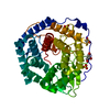 2zzrS S: Starting model for refinement |
|---|---|
| Similar structure data |


-Links
 PDBj
PDBj
- Assembly
| Deposited unit | 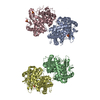
| ||||||||
|---|---|---|---|---|---|---|---|---|---|
| 1 | 
| ||||||||
| 2 | 
| ||||||||
| 3 | 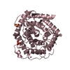
| ||||||||
| 4 | 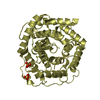
| ||||||||
| 5 | 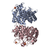
| ||||||||
| 6 | 
| ||||||||
| Unit cell |
|
-Components
| #1: Protein | Mass: 46640.695 Da / Num. of mol.: 4 / Mutation: D115N Source method: isolated from a genetically manipulated source Source: (gene. exp.) Streptococcus agalactiae (bacteria) / Strain: NEM316 / Gene: gbs1889 / Plasmid: pET21B / Production host: References: UniProt: Q8E372, Hydrolases; Glycosylases; Glycosidases, i.e. enzymes that hydrolyse O- and S-glycosyl compounds #2: Chemical | ChemComp-SO4 /   Mass: 96.063 Da / Num. of mol.: 6 / Source method: obtained synthetically / Formula: SO4 Mass: 96.063 Da / Num. of mol.: 6 / Source method: obtained synthetically / Formula: SO4#3: Water | ChemComp-HOH / |  Mass: 18.015 Da / Num. of mol.: 691 / Source method: isolated from a natural source / Formula: H2O Mass: 18.015 Da / Num. of mol.: 691 / Source method: isolated from a natural source / Formula: H2O |
|---|
-Experimental details
-Experiment
| Experiment | Method: X-RAY DIFFRACTION / Number of used crystals: 1 |
|---|
- Sample preparation
| Crystal | Density Matthews: 2.24 Å3/Da / Density % sol: 45.13 % |
|---|---|
| Crystal grow | Temperature: 293 K / Method: vapor diffusion, sitting drop / pH: 9 Details: 1.5M ammonium sulphate, 0.1M Tris-HCl, pH 9.0, VAPOR DIFFUSION, SITTING DROP, temperature 293K |
-Data collection
| Diffraction | Mean temperature: 100 K |
|---|---|
| Diffraction source | Source: SYNCHROTRON / Site: SPring-8  / Beamline: BL38B1 / Wavelength: 1 Å / Beamline: BL38B1 / Wavelength: 1 Å |
| Detector | Type: ADSC QUANTUM 210 / Detector: CCD / Date: May 22, 2009 |
| Radiation | Monochromator: Fixed exit Si(111) double crystal monochromato Protocol: SINGLE WAVELENGTH / Monochromatic (M) / Laue (L): M / Scattering type: x-ray |
| Radiation wavelength | Wavelength: 1 Å / Relative weight: 1 |
| Reflection | Resolution: 1.87→50 Å / Num. all: 136222 / Num. obs: 133399 / % possible obs: 97.9 % / Observed criterion σ(F): 0 / Observed criterion σ(I): 0 / Redundancy: 4.7 % / Rmerge(I) obs: 0.046 / Net I/σ(I): 38.8 |
| Reflection shell | Resolution: 1.87→1.94 Å / Redundancy: 4.4 % / Rmerge(I) obs: 0.336 / Num. unique all: 13014 / % possible all: 95.8 |
- Processing
| Software |
| ||||||||||||||||||||||||||||||||||||||||||||||||||||||||||||||||||||||||||||||||||||||||||||||||||||||||||||||||||||||||||||||||||||||||||||||||||||||||||||||||||||||||||
|---|---|---|---|---|---|---|---|---|---|---|---|---|---|---|---|---|---|---|---|---|---|---|---|---|---|---|---|---|---|---|---|---|---|---|---|---|---|---|---|---|---|---|---|---|---|---|---|---|---|---|---|---|---|---|---|---|---|---|---|---|---|---|---|---|---|---|---|---|---|---|---|---|---|---|---|---|---|---|---|---|---|---|---|---|---|---|---|---|---|---|---|---|---|---|---|---|---|---|---|---|---|---|---|---|---|---|---|---|---|---|---|---|---|---|---|---|---|---|---|---|---|---|---|---|---|---|---|---|---|---|---|---|---|---|---|---|---|---|---|---|---|---|---|---|---|---|---|---|---|---|---|---|---|---|---|---|---|---|---|---|---|---|---|---|---|---|---|---|---|---|---|
| Refinement | Method to determine structure: MOLECULAR REPLACEMENT Starting model: PDB ENTRY 2ZZR Resolution: 2→27.48 Å / Cor.coef. Fo:Fc: 0.952 / Cor.coef. Fo:Fc free: 0.932 / SU B: 3.588 / SU ML: 0.102 / Cross valid method: THROUGHOUT / σ(F): 0 / ESU R: 0.187 / ESU R Free: 0.159 / Stereochemistry target values: MAXIMUM LIKELIHOOD Details: HYDROGENS HAVE BEEN ADDED IN THE RIDING POSITIONS U VALUES: REFINED INDIVIDUALLY
| ||||||||||||||||||||||||||||||||||||||||||||||||||||||||||||||||||||||||||||||||||||||||||||||||||||||||||||||||||||||||||||||||||||||||||||||||||||||||||||||||||||||||||
| Solvent computation | Ion probe radii: 0.8 Å / Shrinkage radii: 0.8 Å / VDW probe radii: 1.4 Å / Solvent model: MASK | ||||||||||||||||||||||||||||||||||||||||||||||||||||||||||||||||||||||||||||||||||||||||||||||||||||||||||||||||||||||||||||||||||||||||||||||||||||||||||||||||||||||||||
| Displacement parameters | Biso mean: 24.432 Å2
| ||||||||||||||||||||||||||||||||||||||||||||||||||||||||||||||||||||||||||||||||||||||||||||||||||||||||||||||||||||||||||||||||||||||||||||||||||||||||||||||||||||||||||
| Refinement step | Cycle: LAST / Resolution: 2→27.48 Å
| ||||||||||||||||||||||||||||||||||||||||||||||||||||||||||||||||||||||||||||||||||||||||||||||||||||||||||||||||||||||||||||||||||||||||||||||||||||||||||||||||||||||||||
| Refine LS restraints |
| ||||||||||||||||||||||||||||||||||||||||||||||||||||||||||||||||||||||||||||||||||||||||||||||||||||||||||||||||||||||||||||||||||||||||||||||||||||||||||||||||||||||||||
| LS refinement shell | Resolution: 2→2.052 Å / Total num. of bins used: 20
|
