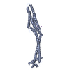
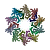
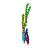
Journal: Nat Commun / Year: 2018 Title: Structure and mechanism of the two-component α-helical pore-forming toxin YaxAB. Authors: Bastian Bräuning / Eva Bertosin / Florian Praetorius / Christian Ihling / Alexandra Schatt / Agnes Adler / Klaus Richter / Andrea Sinz / Hendrik Dietz / Michael Groll / Abstract: Pore-forming toxins (PFT) are virulence factors that transform from soluble to membrane-bound states. The Yersinia YaxAB system represents a family of binary α-PFTs with orthologues in human, ...Pore-forming toxins (PFT) are virulence factors that transform from soluble to membrane-bound states. The Yersinia YaxAB system represents a family of binary α-PFTs with orthologues in human, insect, and plant pathogens, with unknown structures. YaxAB was shown to be cytotoxic and likely involved in pathogenesis, though the molecular basis for its two-component lytic mechanism remains elusive. Here, we present crystal structures of YaxA and YaxB, together with a cryo-electron microscopy map of the YaxAB complex. Our structures reveal a pore predominantly composed of decamers of YaxA-YaxB heterodimers. Both subunits bear membrane-active moieties, but only YaxA is capable of binding to membranes by itself. YaxB can subsequently be recruited to membrane-associated YaxA and induced to present its lytic transmembrane helices. Pore formation can progress by further oligomerization of YaxA-YaxB dimers. Our results allow for a comparison between pore assemblies belonging to the wider ClyA-like family of α-PFTs, highlighting diverse pore architectures.
Resolution: 4→48.82 Å / Cor.coef. Fo:Fc: 0.901 / Cor.coef. Fo:Fc free: 0.928 / SU B: 155.607 / SU ML: 0.904 / Cross valid method: THROUGHOUT / ESU R Free: 0.847 / Stereochemistry target values: MAXIMUM LIKELIHOOD / Details: HYDROGENS HAVE BEEN ADDED IN THE RIDING POSITIONS
Rfactor
Num. reflection
% reflection
Selection details
Rfree
0.33881
282
5 %
RANDOM
Rwork
0.32555
-
-
-
obs
0.32625
5351
98.65 %
-
Solvent computation
Ion probe radii: 0.8 Å / Shrinkage radii: 0.8 Å / VDW probe radii: 1.2 Å / Solvent model: MASK
 Movie
Movie Controller


 Open data
 Basic information
 Components
 Keywords
 Function and homology information
 Yersinia enterocolitica (bacteria)
 X-RAY DIFFRACTION /
  Authors
 Citation
 Structure visualization
 Downloads & links
 Other downloads
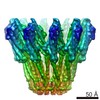


 PDBj
PDBj Assembly
 Sample preparation
 / Beamline: X06SA / Wavelength: 1 Å
 Processing