 Movie
Movie Controller

[English] 日本語
 Yorodumi
- PDB-6ejv: Nuclease NucB from Bacillus licheniformis in sulphate free conditions -
+ Open data

- Basic information
| Entry | Database: PDB / ID: 6ejv | ||||||||||||
|---|---|---|---|---|---|---|---|---|---|---|---|---|---|
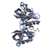
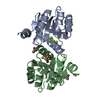
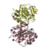
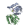
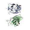
| Title | Nuclease NucB from Bacillus licheniformis in sulphate free conditions | ||||||||||||
 Components Components | Nuclease | ||||||||||||
 Keywords Keywords | HYDROLASE / nuclease / DNAse / metal dependent | ||||||||||||
| Function / homology | Deoxyribonuclease NucA/NucB / Deoxyribonuclease NucA/NucB / Nuclease Function and homology information Function and homology information | ||||||||||||
| Biological species |  | ||||||||||||
| Method |  X-RAY DIFFRACTION / MOLECULAR REPLACEMENT / Resolution: 1.75 Å X-RAY DIFFRACTION / MOLECULAR REPLACEMENT / Resolution: 1.75 Å | ||||||||||||
 Authors Authors | Stransky, J. / Dohnalek, J. / Oestergaard, L.A. | ||||||||||||
| Funding support |  Czech Republic, 3items Czech Republic, 3items
| ||||||||||||
 Citation Citation | Journal: To Be Published Title: Structure of novel nuclease NucB from Bacillus licheniformis Authors: Stransky, J. / Dohnalek, J. / Oestergaard, L.H. | ||||||||||||
| History |
|
- Structure visualization
| Structure viewer | Molecule: MolmilJmol/JSmol |
|---|
- Downloads & links
-Download
| PDBx/mmCIF format | 6ejv.cif.gz | 65 KB | Display | PDBx/mmCIF format |
|---|---|---|---|---|
| PDB format | pdb6ejv.ent.gz | 46.2 KB | Display | PDB format |
| PDBx/mmJSON format | 6ejv.json.gz | Tree view | PDBx/mmJSON format | |
| Others |  Other downloads Other downloads |
-Validation report
| Arichive directory | https://data.pdbj.org/pub/pdb/validation_reports/ej/6ejvftp://data.pdbj.org/pub/pdb/validation_reports/ej/6ejv | HTTPS FTP |
|---|
-Related structure data
| Related structure data | 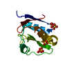 6ejsSC 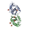 6ejtC 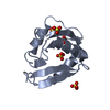 6ejuC  9fj7C C: citing same article ( S: Starting model for refinement |
|---|---|
| Similar structure data |
-Links
 PDBj
PDBj- Assembly
| Deposited unit | 
| ||||||||
|---|---|---|---|---|---|---|---|---|---|
| 1 |
| ||||||||
| Unit cell |
|
-Components
| #1: Protein | Mass: 12001.333 Da / Num. of mol.: 2 Source method: isolated from a genetically manipulated source Details: putative EC3.1.21.1 / Source: (gene. exp.) #2: Chemical | ChemComp-TRS / |   Mass: 122.143 Da / Num. of mol.: 1 / Source method: obtained synthetically / Formula: C4H12NO3 / Comment: pH buffer*YM Mass: 122.143 Da / Num. of mol.: 1 / Source method: obtained synthetically / Formula: C4H12NO3 / Comment: pH buffer*YM#3: Water | ChemComp-HOH / |  Mass: 18.015 Da / Num. of mol.: 280 / Source method: isolated from a natural source / Formula: H2O Mass: 18.015 Da / Num. of mol.: 280 / Source method: isolated from a natural source / Formula: H2OHas protein modification | Y | |
|---|
-Experimental details
-Experiment
| Experiment | Method: X-RAY DIFFRACTION / Number of used crystals: 1 |
|---|
- Sample preparation
| Crystal | Density Matthews: 2.5 Å3/Da / Density % sol: 50.6 % |
|---|---|
| Crystal grow | Temperature: 293 K / Method: vapor diffusion, sitting drop / pH: 8.5 Details: 0.12 M 1,6-Hexanediol 0.12 M 1-Butanol 0.12 M 1,2-Propanediol 0.12 M 2-Propanol 0.12 M 1,4-Butanediol 0.12 M 1,3-Propanediol 0.1 M Tris and Bicine pH 8.5 12.5 % v/v 2-Methyl- -2,4,- ...Details: 0.12 M 1,6-Hexanediol 0.12 M 1-Butanol 0.12 M 1,2-Propanediol 0.12 M 2-Propanol 0.12 M 1,4-Butanediol 0.12 M 1,3-Propanediol 0.1 M Tris and Bicine pH 8.5 12.5 % v/v 2-Methyl- -2,4,-pentanediol 12.5 % PEG 1000 12.5 % w/v PEG 3350 |
-Data collection
| Diffraction | Mean temperature: 110 K |
|---|---|
| Diffraction source | Source: LIQUID ANODE / Type: Excillum MetalJet D2+ 70 kV / Wavelength: 1.3418 Å |
| Detector | Type: Bruker PHOTON II / Detector: PIXEL / Date: May 5, 2017 / Details: HELIOS MX |
| Radiation | Monochromator: GOEBLE MIRRORS / Protocol: SINGLE WAVELENGTH / Monochromatic (M) / Laue (L): M / Scattering type: x-ray |
| Radiation wavelength | Wavelength: 1.3418 Å / Relative weight: 1 |
| Reflection | Resolution: 1.75→45.5 Å / Num. obs: 24852 / % possible obs: 99.9 % / Observed criterion σ(I): -3 / Redundancy: 9.1 % / Biso Wilson estimate: 14.48 Å2 / CC1/2: 0.998 / Rmerge(I) obs: 0.09 / Rpim(I) all: 0.045 / Rrim(I) all: 0.101 / Net I/σ(I): 17.9 |
| Reflection shell | Resolution: 1.75→1.78 Å / Redundancy: 7.1 % / Rmerge(I) obs: 0.872 / Mean I/σ(I) obs: 1.8 / Num. unique obs: 1354 / CC1/2: 0.78 / Rpim(I) all: 0.518 / Rrim(I) all: 1.018 / % possible all: 100 |
- Processing
| Software |
| |||||||||||||||||||||||||||||||||||||||||||||||||||||||||||||||||||||||||||
|---|---|---|---|---|---|---|---|---|---|---|---|---|---|---|---|---|---|---|---|---|---|---|---|---|---|---|---|---|---|---|---|---|---|---|---|---|---|---|---|---|---|---|---|---|---|---|---|---|---|---|---|---|---|---|---|---|---|---|---|---|---|---|---|---|---|---|---|---|---|---|---|---|---|---|---|---|
| Refinement | Method to determine structure: MOLECULAR REPLACEMENT Starting model: 6EJS Resolution: 1.75→45.5 Å / Cor.coef. Fo:Fc: 0.964 / Cor.coef. Fo:Fc free: 0.961 / Cross valid method: THROUGHOUT / σ(F): 0 / ESU R: 0.102 / ESU R Free: 0.087 Details: HYDROGENS HAVE BEEN ADDED IN THE RIDING POSITIONS U VALUES : REFINED INDIVIDUALLY
| |||||||||||||||||||||||||||||||||||||||||||||||||||||||||||||||||||||||||||
| Solvent computation | Ion probe radii: 0.8 Å / Shrinkage radii: 0.8 Å / VDW probe radii: 1.2 Å | |||||||||||||||||||||||||||||||||||||||||||||||||||||||||||||||||||||||||||
| Displacement parameters | Biso max: 71.45 Å2 / Biso mean: 22.3148 Å2 / Biso min: 11.17 Å2
| |||||||||||||||||||||||||||||||||||||||||||||||||||||||||||||||||||||||||||
| Refinement step | Cycle: LAST / Resolution: 1.75→45.5 Å
| |||||||||||||||||||||||||||||||||||||||||||||||||||||||||||||||||||||||||||
| Refine LS restraints |
| |||||||||||||||||||||||||||||||||||||||||||||||||||||||||||||||||||||||||||
| LS refinement shell | Resolution: 1.75→1.795 Å / Total num. of bins used: 20
|
