Monochromator: double crystal / Protocol: SINGLE WAVELENGTH / Monochromatic (M) / Laue (L): M / Scattering type: x-ray
Radiation wavelength
Wavelength: 0.97926 Å / Relative weight: 1
Reflection
Resolution: 2.094→35 Å / Num. all: 15323 / Num. obs: 15260 / % possible obs: 99.6 % / Observed criterion σ(I): -3 / Redundancy: 6.6 % / Biso Wilson estimate: 27.4 Å2 / Rmerge(I) obs: 0.138 / Net I/σ(I): 16.6
Reflection shell
Resolution: 2.1→2.14 Å / Redundancy: 6.3 % / Rmerge(I) obs: 0.743 / Mean I/σ(I) obs: 2.4 / Num. unique all: 749 / % possible all: 100
-
Processing
Software
Name
Version
Classification
SBC-Collect
datacollection
SHELX
modelbuilding
MLPHARE
phasing
DM
modelbuilding
ARP/wARP
modelbuilding
Coot
modelbuilding
PHENIX
(phenix.refine: 1.6.1_357)
refinement
HKL-3000
datareduction
HKL-3000
datascaling
SHELX
phasing
DM
phasing
Refinement
Method to determine structure: SAD / Resolution: 2.094→30.202 Å / SU ML: 0.24 / Cross valid method: THROUGHOUT / σ(F): 1.34 / Stereochemistry target values: ML
Rfactor
Num. reflection
% reflection
Selection details
Rfree
0.2268
881
5.79 %
random
Rwork
0.1772
-
-
-
all
0.1799
15213
-
-
obs
0.1799
15213
99.44 %
-
Solvent computation
Shrinkage radii: 0.9 Å / VDW probe radii: 1.11 Å / Solvent model: FLAT BULK SOLVENT MODEL / Bsol: 42.704 Å2 / ksol: 0.424 e/Å3
Displacement parameters
Baniso -1
Baniso -2
Baniso -3
1-
5.9105 Å2
0 Å2
-0 Å2
2-
-
5.9105 Å2
0 Å2
3-
-
-
-11.8211 Å2
Refinement step
Cycle: LAST / Resolution: 2.094→30.202 Å
Protein
Nucleic acid
Ligand
Solvent
Total
Num. atoms
1885
0
12
53
1950
Refine LS restraints
Refine-ID
Type
Dev ideal
Number
X-RAY DIFFRACTION
f_bond_d
0.007
1959
X-RAY DIFFRACTION
f_angle_d
1
2634
X-RAY DIFFRACTION
f_dihedral_angle_d
16.357
728
X-RAY DIFFRACTION
f_chiral_restr
0.067
271
X-RAY DIFFRACTION
f_plane_restr
0.003
344
LS refinement shell
Resolution (Å)
Rfactor Rfree
Num. reflection Rfree
Rfactor Rwork
Num. reflection Rwork
Refine-ID
% reflection obs (%)
2.0942-2.2253
0.2711
164
0.2237
2296
X-RAY DIFFRACTION
99
2.2253-2.3971
0.2614
153
0.2055
2337
X-RAY DIFFRACTION
100
2.3971-2.6382
0.2439
146
0.1896
2368
X-RAY DIFFRACTION
100
2.6382-3.0196
0.2296
147
0.1804
2385
X-RAY DIFFRACTION
100
3.0196-3.8032
0.2224
121
0.155
2435
X-RAY DIFFRACTION
100
3.8032-30.2054
0.2034
150
0.1693
2511
X-RAY DIFFRACTION
98
Refinement TLS params.
Method: refined / Refine-ID: X-RAY DIFFRACTION
ID
L11 (°2)
L12 (°2)
L13 (°2)
L22 (°2)
L23 (°2)
L33 (°2)
S11 (Å °)
S12 (Å °)
S13 (Å °)
S21 (Å °)
S22 (Å °)
S23 (Å °)
S31 (Å °)
S32 (Å °)
S33 (Å °)
T11 (Å2)
T12 (Å2)
T13 (Å2)
T22 (Å2)
T23 (Å2)
T33 (Å2)
Origin x (Å)
Origin y (Å)
Origin z (Å)
1
2.9263
-1.7418
1.5687
1.6905
0.913
7.249
-0.3959
-0.0536
1.437
0.4844
0.2824
2.0261
-0.5854
-1.1594
-0.1999
0.4669
-0.0029
0.151
0.4931
-0.0259
0.7329
53.6192
36.2857
20.1468
2
0.7745
0.8798
-0.1682
2.0646
1.5272
3.047
-0.0438
0.0906
0.006
-0.1536
0.0159
0.1459
-0.0466
-0.1466
-0.003
0.2528
0.0157
-0.0175
0.2505
0.0461
0.2613
55.486
19.9776
5.3244
3
9.8557
5.335
5.3961
4.397
2.3787
3.1659
0.6339
0.189
-0.0076
0.5583
0.0755
-0.1335
-0.4475
-1.0952
-0.0313
0.6507
-0.082
0.0451
0.6365
0.054
0.5786
46.7916
6.6911
14.4851
4
0.3106
-0.0631
-0.5058
1.4435
-0.6215
3.3104
-0.16
-0.1302
-0.1302
-0.0346
0.227
0.0196
0.8128
-0.232
0.0565
0.3509
-0.0744
-0.0333
0.2707
0.0336
0.2109
54.6835
13.5804
7.8448
5
5.3739
-3.5558
-6.995
8.4278
2.7465
9.9747
0.2909
-0.9312
0.8176
0.699
0.2352
-0.8345
-0.9054
0.7584
-0.2898
0.2607
-0.0861
-0.0608
0.3355
0.0586
0.2477
60.5653
14.3628
11.2961
6
3.0464
3.0846
1.3525
3.2792
1.9108
2.6274
0.2022
0.6751
-0.6223
-0.523
0.134
-1.0269
0.3662
1.9837
-1.3223
0.2123
0.0757
0.0434
0.5136
-0.0276
0.3586
68.5957
22.5546
52.1667
7
2.5796
0.3823
0.3471
0.1388
0.5284
1.5559
0.0139
-0.1662
-0.1988
0.0739
0.0298
-0.0436
-0.0289
-0.0848
-0.0064
0.2
-0.002
0.0055
0.205
0.0343
0.2089
50.4217
18.2834
51.594
8
2.4785
1.4823
-1.1688
3.1913
-1.5288
1.9595
-0.0514
0.6222
-0.0587
-0.3194
0.176
0.0593
-0.0706
-0.073
0.0471
0.3401
-0.0563
0.0023
0.4596
-0.0176
0.2522
42.0752
18.1603
36.8427
9
3.5111
1.4604
-0.9298
2.8811
-3.2958
3.9702
-0.0427
-0.1008
0.0923
0.1945
-0.043
0.1095
-0.1367
0.007
0.064
0.1811
0.035
-0.0302
0.2465
0.0237
0.2303
38.0104
21.9464
47.8752
10
9.0464
-3.1814
-5.1183
4.0911
2.5326
3.9818
0.3063
0.07
0.3821
-0.1285
0.0133
-0.2429
-0.1907
0.0681
-0.1216
0.2395
-0.026
-0.0289
0.2095
0.0141
0.1486
53.2395
21.6072
41.0488
Refinement TLS group
ID
Refine-ID
Refine TLS-ID
Selection details
1
X-RAY DIFFRACTION
1
(chainAandresid4:11)
2
X-RAY DIFFRACTION
2
(chainAandresid12:66)
3
X-RAY DIFFRACTION
3
(chainAandresid67:75)
4
X-RAY DIFFRACTION
4
(chainAandresid76:106)
5
X-RAY DIFFRACTION
5
(chainAandresid107:114)
6
X-RAY DIFFRACTION
6
(chainBandresid5:12)
7
X-RAY DIFFRACTION
7
(chainBandresid13:55)
8
X-RAY DIFFRACTION
8
(chainBandresid56:75)
9
X-RAY DIFFRACTION
9
(chainBandresid76:94)
10
X-RAY DIFFRACTION
10
(chainBandresid95:114)
+
About Yorodumi
-
News
-
Feb 9, 2022. New format data for meta-information of EMDB entries
New format data for meta-information of EMDB entries
Version 3 of the EMDB header file is now the official format.
The previous official version 1.9 will be removed from the archive.
In the structure databanks used in Yorodumi, some data are registered as the other names, "COVID-19 virus" and "2019-nCoV". Here are the details of the virus and the list of structure data.
Jan 31, 2019. EMDB accession codes are about to change! (news from PDBe EMDB page)
EMDB accession codes are about to change! (news from PDBe EMDB page)
The allocation of 4 digits for EMDB accession codes will soon come to an end. Whilst these codes will remain in use, new EMDB accession codes will include an additional digit and will expand incrementally as the available range of codes is exhausted. The current 4-digit format prefixed with “EMD-” (i.e. EMD-XXXX) will advance to a 5-digit format (i.e. EMD-XXXXX), and so on. It is currently estimated that the 4-digit codes will be depleted around Spring 2019, at which point the 5-digit format will come into force.
The EM Navigator/Yorodumi systems omit the EMD- prefix.
Related info.:Q: What is EMD? / ID/Accession-code notation in Yorodumi/EM Navigator
Yorodumi is a browser for structure data from EMDB, PDB, SASBDB, etc.
This page is also the successor to EM Navigator detail page, and also detail information page/front-end page for Omokage search.
The word "yorodu" (or yorozu) is an old Japanese word meaning "ten thousand". "mi" (miru) is to see.
Related info.:EMDB / PDB / SASBDB / Comparison of 3 databanks / Yorodumi Search / Aug 31, 2016. New EM Navigator & Yorodumi / Yorodumi Papers / Jmol/JSmol / Function and homology information / Changes in new EM Navigator and Yorodumi
 Movie
Movie Controller

 Yorodumi
 Open data
 Basic information
 Components
 Keywords
 Function and homology information
 Nostoc sp. (bacteria)
 X-RAY DIFFRACTION /
  Authors
 Citation
 Structure visualization
 Downloads & links
 Other downloads

 PDBj
PDBj


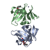
 Assembly
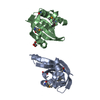

 Mass: 92.094 Da / Num. of mol.: 2 / Source method: obtained synthetically / Formula: C3H8O3
 Mass: 18.015 Da / Num. of mol.: 53 / Source method: isolated from a natural source / Formula: H2O
 Sample preparation
 / Beamline: 19-BM / Wavelength: 0.97926 Å
 Processing