 Movie
Movie Controller


+ Open data

- Basic information
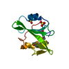
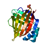
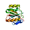
| Entry | Database: PDB / ID: 6ejs | ||||||||||||
|---|---|---|---|---|---|---|---|---|---|---|---|---|---|
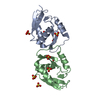
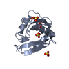
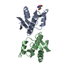
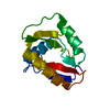
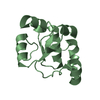
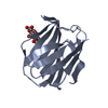
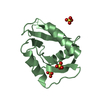
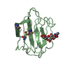
| Title | Nuclease NucB from Bacillus licheniformis in P212121 space group | ||||||||||||
 Components Components | Nuclease | ||||||||||||
 Keywords Keywords | HYDROLASE / nuclease / DNAse / metal dependent | ||||||||||||
| Function / homology | Deoxyribonuclease NucA/NucB / Deoxyribonuclease NucA/NucB / Nuclease Function and homology information Function and homology information | ||||||||||||
| Biological species |  | ||||||||||||
| Method |  X-RAY DIFFRACTION / SYNCHROTRON / SAD / Resolution: 1.6 Å X-RAY DIFFRACTION / SYNCHROTRON / SAD / Resolution: 1.6 Å | ||||||||||||
 Authors Authors | Stransky, J. / Dohnalek, J. / Oestergaard, L.A. | ||||||||||||
| Funding support |  Czech Republic, 3items Czech Republic, 3items
| ||||||||||||
 Citation Citation | Journal: To Be Published Title: Structure of novel nuclease NucB from Bacillus licheniformis Authors: Stransky, J. / Dohnalek, J. / Oestergaard, L.H. | ||||||||||||
| History |
|
- Structure visualization
| Structure viewer | Molecule: MolmilJmol/JSmol |
|---|
- Downloads & links
-Download
| PDBx/mmCIF format | 6ejs.cif.gz | 41.3 KB | Display | PDBx/mmCIF format |
|---|---|---|---|---|
| PDB format | pdb6ejs.ent.gz | 26.8 KB | Display | PDB format |
| PDBx/mmJSON format | 6ejs.json.gz | Tree view | PDBx/mmJSON format | |
| Others |  Other downloads Other downloads |
-Validation report
| Arichive directory | https://data.pdbj.org/pub/pdb/validation_reports/ej/6ejsftp://data.pdbj.org/pub/pdb/validation_reports/ej/6ejs | HTTPS FTP |
|---|
-Related structure data


-Links
 PDBj
PDBj- Assembly
| Deposited unit | 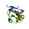
| ||||||||
|---|---|---|---|---|---|---|---|---|---|
| 1 |
| ||||||||
| Unit cell |
|
-Components
| #1: Protein | Mass: 12001.333 Da / Num. of mol.: 1 Source method: isolated from a genetically manipulated source Details: putative EC 3.1.21.1 / Source: (gene. exp.) | ||||
|---|---|---|---|---|---|
| #2: Chemical |   Mass: 96.063 Da / Num. of mol.: 3 / Source method: obtained synthetically / Formula: SO4 Mass: 96.063 Da / Num. of mol.: 3 / Source method: obtained synthetically / Formula: SO4#3: Water | ChemComp-HOH / |  Mass: 18.015 Da / Num. of mol.: 139 / Source method: isolated from a natural source / Formula: H2O Mass: 18.015 Da / Num. of mol.: 139 / Source method: isolated from a natural source / Formula: H2OHas protein modification | Y | |
-Experimental details
-Experiment
| Experiment | Method: X-RAY DIFFRACTION / Number of used crystals: 1 |
|---|
- Sample preparation
| Crystal | Density Matthews: 1.8 Å3/Da / Density % sol: 32 % Preparation: SAD phase problem was solved with the data collected at 1.9 A wavelength, while the presented structure is refined against the 0.9184 A dataset |
|---|---|
| Crystal grow | Temperature: 291 K / Method: vapor diffusion, hanging drop / pH: 8.5 / Details: 2.2 M ammonium sulphate, 0.1 M Tris pH 8.5 |
-Data collection
| Diffraction | Mean temperature: 100 K | |||||||||
|---|---|---|---|---|---|---|---|---|---|---|
| Diffraction source | Source: SYNCHROTRON / Site: BESSY  / Beamline: 14.2 / Wavelength: 1.9,0.9184 / Beamline: 14.2 / Wavelength: 1.9,0.9184 | |||||||||
| Detector | Type: MARMOSAIC 225 mm CCD / Detector: CCD / Date: Nov 15, 2012 | |||||||||
| Radiation | Monochromator: 2 MIRROR AND DOUBLE-CRYSTAL MONOCHROMATOR / Protocol: SINGLE WAVELENGTH / Monochromatic (M) / Laue (L): M / Scattering type: x-ray | |||||||||
| Radiation wavelength |
| |||||||||
| Reflection | Resolution: 1.6→47.41 Å / Num. obs: 10804 / % possible obs: 90.1 % / Observed criterion σ(I): -3 / Redundancy: 6.7 % / Biso Wilson estimate: 13 Å2 Details: SAD phase problem was solved with the data collected at 1.9 A wavelength, while the presented structure is refined against the 0.9184 A dataset CC1/2: 0.999 / Rmerge(I) obs: 0.037 / Rpim(I) all: 0.022 / Rrim(I) all: 0.043 / Net I/σ(I): 29.8 | |||||||||
| Reflection shell | Resolution: 1.6→1.63 Å / Redundancy: 5.9 % / Rmerge(I) obs: 0.116 / Num. unique obs: 448 / Rpim(I) all: 0.079 / Rrim(I) all: 0.142 / % possible all: 77.7 |
- Processing
| Software |
| |||||||||||||||||||||||||||||||||||||||||||||||||||||||||||||||||||||||||||
|---|---|---|---|---|---|---|---|---|---|---|---|---|---|---|---|---|---|---|---|---|---|---|---|---|---|---|---|---|---|---|---|---|---|---|---|---|---|---|---|---|---|---|---|---|---|---|---|---|---|---|---|---|---|---|---|---|---|---|---|---|---|---|---|---|---|---|---|---|---|---|---|---|---|---|---|---|
| Refinement | Method to determine structure: SAD / Resolution: 1.6→36.28 Å / Cor.coef. Fo:Fc: 0.953 / Cor.coef. Fo:Fc free: 0.961 / Cross valid method: THROUGHOUT / σ(F): 0 / ESU R: 0.138 / ESU R Free: 0.104 Stereochemistry target values: CCP4 geometric library, version 4.11 Details: HYDROGENS HAVE BEEN ADDED IN THE RIDING POSITIONS U VALUES : REFINED INDIVIDUALLY
| |||||||||||||||||||||||||||||||||||||||||||||||||||||||||||||||||||||||||||
| Solvent computation | Ion probe radii: 0.8 Å / Shrinkage radii: 0.8 Å / VDW probe radii: 1.2 Å | |||||||||||||||||||||||||||||||||||||||||||||||||||||||||||||||||||||||||||
| Displacement parameters | Biso max: 52.61 Å2 / Biso mean: 17.7017 Å2 / Biso min: 8.5 Å2
| |||||||||||||||||||||||||||||||||||||||||||||||||||||||||||||||||||||||||||
| Refinement step | Cycle: LAST / Resolution: 1.6→36.28 Å
| |||||||||||||||||||||||||||||||||||||||||||||||||||||||||||||||||||||||||||
| Refine LS restraints |
| |||||||||||||||||||||||||||||||||||||||||||||||||||||||||||||||||||||||||||
| LS refinement shell | Resolution: 1.6→1.642 Å / Total num. of bins used: 20
|
