 Movie
Movie Controller


+ Open data

- Basic information
| Entry | Database: PDB / ID: 9fj7 | |||||||||
|---|---|---|---|---|---|---|---|---|---|---|
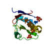
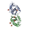
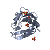
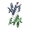
| Title | Nuclease NucB from Bacillus licheniformis mutant N117Q | |||||||||
 Components Components | Nuclease | |||||||||
 Keywords Keywords | DNA BINDING PROTEIN / DNAse / endonuclease / mutant N117Q | |||||||||
| Function / homology | Deoxyribonuclease NucA/NucB / Deoxyribonuclease NucA/NucB / Nuclease Function and homology information Function and homology information | |||||||||
| Biological species |  | |||||||||
| Method |  X-RAY DIFFRACTION / SYNCHROTRON / MOLECULAR REPLACEMENT / Resolution: 1.25 Å X-RAY DIFFRACTION / SYNCHROTRON / MOLECULAR REPLACEMENT / Resolution: 1.25 Å | |||||||||
 Authors Authors | Koval, T. / Stransky, J. / Oestergaard, L.H. / Dohnalek, J. | |||||||||
| Funding support |  Czech Republic, 2items Czech Republic, 2items
| |||||||||
 Citation Citation | Journal: To Be Published Title: Structure of novel nuclease NucB from Bacillus licheniformis Authors: Stransky, J. / Koval, T. / Oestergaard, L.H. / Dohnalek, J. | |||||||||
| History |
|
- Structure visualization
| Structure viewer | Molecule: MolmilJmol/JSmol |
|---|
- Downloads & links
-Download
| PDBx/mmCIF format | 9fj7.cif.gz | 70.6 KB | Display | PDBx/mmCIF format |
|---|---|---|---|---|
| PDB format | pdb9fj7.ent.gz | Display | PDB format | |
| PDBx/mmJSON format | 9fj7.json.gz | Tree view | PDBx/mmJSON format | |
| Others |  Other downloads Other downloads |
-Validation report
| Arichive directory | https://data.pdbj.org/pub/pdb/validation_reports/fj/9fj7ftp://data.pdbj.org/pub/pdb/validation_reports/fj/9fj7 | HTTPS FTP |
|---|
-Related structure data
-Links
 PDBj
PDBj- Assembly
| Deposited unit | 
| ||||||||
|---|---|---|---|---|---|---|---|---|---|
| 1 |
| ||||||||
| Unit cell |
| ||||||||
| Components on special symmetry positions |
|
-Components
| #1: Protein | Mass: 12015.358 Da / Num. of mol.: 1 / Mutation: N117Q Source method: isolated from a genetically manipulated source Source: (gene. exp.) |
|---|---|
| #2: Chemical | ChemComp-MPD / (  Mass: 118.174 Da / Num. of mol.: 1 / Source method: obtained synthetically / Formula: C6H14O2 / Comment: precipitant*YM Mass: 118.174 Da / Num. of mol.: 1 / Source method: obtained synthetically / Formula: C6H14O2 / Comment: precipitant*YM |
| #3: Water | ChemComp-HOH /  Mass: 18.015 Da / Num. of mol.: 222 / Source method: isolated from a natural source / Formula: H2O Mass: 18.015 Da / Num. of mol.: 222 / Source method: isolated from a natural source / Formula: H2O |
| Has ligand of interest | N |
| Has protein modification | Y |
-Experimental details
-Experiment
| Experiment | Method: X-RAY DIFFRACTION / Number of used crystals: 1 |
|---|
- Sample preparation
| Crystal | Density Matthews: 2.92 Å3/Da / Density % sol: 57.9 % |
|---|---|
| Crystal grow | Temperature: 291 K / Method: vapor diffusion, sitting drop / pH: 8.5 Details: protein concentration was 18 mg/ml; storage buffer: 20mM Tris pH 8.0, 100mM NaCl; reservoir solution: 0.1 M 1,6-Hexanediol 0.1 M 1-Butanol 0.1 M 1,2-Propanediol 0.1 M 2-Propanol 0.1 M 1,4- ...Details: protein concentration was 18 mg/ml; storage buffer: 20mM Tris pH 8.0, 100mM NaCl; reservoir solution: 0.1 M 1,6-Hexanediol 0.1 M 1-Butanol 0.1 M 1,2-Propanediol 0.1 M 2-Propanol 0.1 M 1,4-Butanediol 0.1 M 1,3-Propanediol 0.85 M Tris and Bicine pH 8.5 10.5 % v/v 2-Methyl- -2,4,-pentanediol 10.5 % PEG 1000 10.5 % w/v PEG 3350 |
-Data collection
| Diffraction | Mean temperature: 100 K / Serial crystal experiment: N |
|---|---|
| Diffraction source | Source: SYNCHROTRON / Site: BESSY  / Beamline: 14.1 / Wavelength: 0.9184 Å / Beamline: 14.1 / Wavelength: 0.9184 Å |
| Detector | Type: DECTRIS PILATUS 6M / Detector: PIXEL / Date: Oct 14, 2021 |
| Radiation | Protocol: SINGLE WAVELENGTH / Monochromatic (M) / Laue (L): M / Scattering type: x-ray |
| Radiation wavelength | Wavelength: 0.9184 Å / Relative weight: 1 |
| Reflection | Resolution: 1.25→46.33 Å / Num. obs: 39455 / % possible obs: 100 % / Redundancy: 9.5 % / Biso Wilson estimate: 12 Å2 / CC1/2: 0.998 / Rmerge(I) obs: 0.067 / Rpim(I) all: 0.023 / Rrim(I) all: 0.071 / Net I/σ(I): 22.4 |
| Reflection shell | Resolution: 1.25→1.27 Å / Redundancy: 9.7 % / Rmerge(I) obs: 1.659 / Mean I/σ(I) obs: 1.7 / Num. unique obs: 1926 / CC1/2: 0.659 / Rpim(I) all: 0.556 / Rrim(I) all: 1.752 / % possible all: 100 |
- Processing
| Software |
| |||||||||||||||||||||||||||||||||||||||||||||||||||||||||||||||||||||||||||||||||||||||||||||||||||||||||||||||||||||||||||||||||||||||||||||||||||||||||||
|---|---|---|---|---|---|---|---|---|---|---|---|---|---|---|---|---|---|---|---|---|---|---|---|---|---|---|---|---|---|---|---|---|---|---|---|---|---|---|---|---|---|---|---|---|---|---|---|---|---|---|---|---|---|---|---|---|---|---|---|---|---|---|---|---|---|---|---|---|---|---|---|---|---|---|---|---|---|---|---|---|---|---|---|---|---|---|---|---|---|---|---|---|---|---|---|---|---|---|---|---|---|---|---|---|---|---|---|---|---|---|---|---|---|---|---|---|---|---|---|---|---|---|---|---|---|---|---|---|---|---|---|---|---|---|---|---|---|---|---|---|---|---|---|---|---|---|---|---|---|---|---|---|---|---|---|---|
| Refinement | Method to determine structure: MOLECULAR REPLACEMENT / Resolution: 1.25→46.33 Å / Cor.coef. Fo:Fc: 0.977 / SU B: 1.132 / SU ML: 0.021 / Cross valid method: THROUGHOUT / ESU R: 0.033 Details: Hydrogens have been added in their riding positions. The last reciprocal space refinement was performed against all measured reflections.
| |||||||||||||||||||||||||||||||||||||||||||||||||||||||||||||||||||||||||||||||||||||||||||||||||||||||||||||||||||||||||||||||||||||||||||||||||||||||||||
| Solvent computation | Ion probe radii: 0.8 Å / Shrinkage radii: 0.8 Å / VDW probe radii: 1.2 Å / Solvent model: MASK BULK SOLVENT | |||||||||||||||||||||||||||||||||||||||||||||||||||||||||||||||||||||||||||||||||||||||||||||||||||||||||||||||||||||||||||||||||||||||||||||||||||||||||||
| Displacement parameters | Biso mean: 17.64 Å2
| |||||||||||||||||||||||||||||||||||||||||||||||||||||||||||||||||||||||||||||||||||||||||||||||||||||||||||||||||||||||||||||||||||||||||||||||||||||||||||
| Refinement step | Cycle: LAST / Resolution: 1.25→46.33 Å
| |||||||||||||||||||||||||||||||||||||||||||||||||||||||||||||||||||||||||||||||||||||||||||||||||||||||||||||||||||||||||||||||||||||||||||||||||||||||||||
| Refine LS restraints |
| |||||||||||||||||||||||||||||||||||||||||||||||||||||||||||||||||||||||||||||||||||||||||||||||||||||||||||||||||||||||||||||||||||||||||||||||||||||||||||
| LS refinement shell | Refine-ID: X-RAY DIFFRACTION
|
