 Movie
Movie Controller

[English] 日本語
 Yorodumi
- PDB-6eix: Crystal structure of the kinase domain of the Q207E mutant of ACV... -
+ Open data

- Basic information
| Entry | Database: PDB / ID: 6eix | ||||||
|---|---|---|---|---|---|---|---|
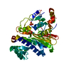
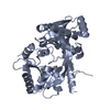
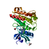
| Title | Crystal structure of the kinase domain of the Q207E mutant of ACVR1 (ALK2) in complex with a 2-aminopyridine inhibitor K02288 | ||||||
 Components Components | Activin receptor type-1 | ||||||
 Keywords Keywords | SIGNALING PROTEIN / Kinase / ALK2 / Receptor / BMP / Structural Genomics / Structural Genomics Consortium / SGC | ||||||
| Function / homology |  Function and homology information Function and homology informationendocardial cushion cell fate commitment / mitral valve morphogenesis / BMP receptor complex / BMP receptor activity / endocardial cushion fusion / cardiac muscle cell fate commitment / atrial septum primum morphogenesis / positive regulation of cardiac epithelial to mesenchymal transition / acute inflammatory response / positive regulation of determination of dorsal identity ...endocardial cushion cell fate commitment / mitral valve morphogenesis / BMP receptor complex / BMP receptor activity / endocardial cushion fusion / cardiac muscle cell fate commitment / atrial septum primum morphogenesis / positive regulation of cardiac epithelial to mesenchymal transition / acute inflammatory response / positive regulation of determination of dorsal identity / transforming growth factor beta receptor activity, type I / smooth muscle cell differentiation / activin receptor activity, type I / activin receptor complex / endocardial cushion formation / pharyngeal system development / receptor protein serine/threonine kinase / activin binding / cellular response to BMP stimulus / transmembrane receptor protein serine/threonine kinase activity / activin receptor signaling pathway / negative regulation of activin receptor signaling pathway / embryonic heart tube morphogenesis / dorsal/ventral pattern formation / gastrulation with mouth forming second / transforming growth factor beta binding / determination of left/right symmetry / atrioventricular valve morphogenesis / neural crest cell migration / branching involved in blood vessel morphogenesis / ventricular septum morphogenesis / negative regulation of G1/S transition of mitotic cell cycle / SMAD binding / germ cell development / mesoderm formation / peptide hormone binding / positive regulation of SMAD protein signal transduction / positive regulation of intracellular signal transduction / regulation of ossification / positive regulation of osteoblast differentiation / negative regulation of signal transduction / positive regulation of bone mineralization / BMP signaling pathway / transforming growth factor beta receptor signaling pathway / protein tyrosine kinase binding / negative regulation of extrinsic apoptotic signaling pathway / cellular response to growth factor stimulus / apical part of cell / osteoblast differentiation / heart development / in utero embryonic development / cell differentiation / protein kinase activity / positive regulation of cell migration / cadherin binding / protein serine/threonine kinase activity / positive regulation of DNA-templated transcription / protein homodimerization activity / positive regulation of transcription by RNA polymerase II / ATP binding / metal ion binding / plasma membrane Similarity search - Function | ||||||
| Biological species |  Homo sapiens (human) Homo sapiens (human) | ||||||
| Method |  X-RAY DIFFRACTION / SYNCHROTRON / MOLECULAR REPLACEMENT / Resolution: 2.3 Å X-RAY DIFFRACTION / SYNCHROTRON / MOLECULAR REPLACEMENT / Resolution: 2.3 Å | ||||||
 Authors Authors | Williams, E.P. / Canning, P. / Sanvitale, C.E. / Krojer, T. / Allerston, C.K. / von Delft, F. / Arrowsmith, C.H. / Edwards, A.M. / Bountra, C. / Bullock, A.N. | ||||||
 Citation Citation | Journal: To be published Title: Crystal structure of the kinase domain of the Q207E mutant of ACVR1 (ALK2) in complex with K02288 Authors: Williams, E.P. / Canning, P. / Sanvitale, C.E. / Bullock, A.N. | ||||||
| History |
|
- Structure visualization
| Structure viewer | Molecule: MolmilJmol/JSmol |
|---|
- Downloads & links
-Download
| PDBx/mmCIF format | 6eix.cif.gz | 139.4 KB | Display | PDBx/mmCIF format |
|---|---|---|---|---|
| PDB format | pdb6eix.ent.gz | 107.7 KB | Display | PDB format |
| PDBx/mmJSON format | 6eix.json.gz | Tree view | PDBx/mmJSON format | |
| Others |  Other downloads Other downloads |
-Validation report
| Arichive directory | https://data.pdbj.org/pub/pdb/validation_reports/ei/6eixftp://data.pdbj.org/pub/pdb/validation_reports/ei/6eix | HTTPS FTP |
|---|
-Related structure data
| Related structure data |  1iasS S: Starting model for refinement |
|---|---|
| Similar structure data |


-Links
 PDBj
PDBj
- Assembly
| Deposited unit | 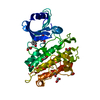
| ||||||||
|---|---|---|---|---|---|---|---|---|---|
| 1 |
| ||||||||
| Unit cell |
|
-Components
| #1: Protein | Mass: 38520.000 Da / Num. of mol.: 1 / Mutation: Q207E Source method: isolated from a genetically manipulated source Details: Missing residues at N and C terminus due to insufficient density to model them. Source: (gene. exp.) Homo sapiens (human) / Gene: ACVR1, ACVRLK2 / Plasmid: pFB-LIC-Bse / Cell line (production host): Sf9 / Production host:   Spodoptera frugiperda (fall armyworm) Spodoptera frugiperda (fall armyworm)References: UniProt: Q04771, receptor protein serine/threonine kinase | ||
|---|---|---|---|
| #2: Chemical | ChemComp-A3F /   Mass: 352.384 Da / Num. of mol.: 1 / Source method: obtained synthetically / Formula: C20H20N2O4 / Feature type: SUBJECT OF INVESTIGATION Mass: 352.384 Da / Num. of mol.: 1 / Source method: obtained synthetically / Formula: C20H20N2O4 / Feature type: SUBJECT OF INVESTIGATION | ||
| #3: Chemical | ChemComp-EDO /   Mass: 62.068 Da / Num. of mol.: 9 / Source method: obtained synthetically / Formula: C2H6O2 Mass: 62.068 Da / Num. of mol.: 9 / Source method: obtained synthetically / Formula: C2H6O2#4: Water | ChemComp-HOH / |  Mass: 18.015 Da / Num. of mol.: 64 / Source method: isolated from a natural source / Formula: H2O Mass: 18.015 Da / Num. of mol.: 64 / Source method: isolated from a natural source / Formula: H2O |
-Experimental details
-Experiment
| Experiment | Method: X-RAY DIFFRACTION / Number of used crystals: 1 |
|---|
- Sample preparation
| Crystal | Density Matthews: 2.21 Å3/Da / Density % sol: 44.35 % |
|---|---|
| Crystal grow | Temperature: 293 K / Method: vapor diffusion, sitting drop / pH: 6.5 / Details: 0.1M MES pH 6.5 -- 12%(w/v) PEG 20000 |
-Data collection
| Diffraction | Mean temperature: 100 K |
|---|---|
| Diffraction source | Source: SYNCHROTRON / Site: Diamond  / Beamline: I03 / Wavelength: 0.9778 Å / Beamline: I03 / Wavelength: 0.9778 Å |
| Detector | Type: DECTRIS PILATUS 6M / Detector: PIXEL / Date: Apr 21, 2012 Details: Kirkpatrick Baez bimorph mirror pair for horizontal and vertical focussing |
| Radiation | Monochromator: Double Crystal / Protocol: SINGLE WAVELENGTH / Monochromatic (M) / Laue (L): M / Scattering type: x-ray |
| Radiation wavelength | Wavelength: 0.9778 Å / Relative weight: 1 |
| Reflection | Resolution: 2.3→19.78 Å / Num. obs: 14813 / % possible obs: 98.9 % / Observed criterion σ(I): 2 / Redundancy: 3 % / CC1/2: 0.984 / Rmerge(I) obs: 0.122 / Rpim(I) all: 0.082 / Rrim(I) all: 0.148 / Net I/σ(I): 5.4 |
| Reflection shell | Resolution: 2.3→2.38 Å / Redundancy: 3.1 % / Rmerge(I) obs: 0.511 / Mean I/σ(I) obs: 2.2 / Num. unique obs: 1444 / CC1/2: 0.749 / Rpim(I) all: 0.344 / Rrim(I) all: 0.619 / % possible all: 99.1 |
- Processing
| Software |
| ||||||||||||||||||||||||||||||||||||||||||||||||||||||||||||||||||||||||||||||||||||||||||||||||||||||||||||||||||||||||||||||||||||||||||||||||||||||||||||||||||||||||||||||||||||||
|---|---|---|---|---|---|---|---|---|---|---|---|---|---|---|---|---|---|---|---|---|---|---|---|---|---|---|---|---|---|---|---|---|---|---|---|---|---|---|---|---|---|---|---|---|---|---|---|---|---|---|---|---|---|---|---|---|---|---|---|---|---|---|---|---|---|---|---|---|---|---|---|---|---|---|---|---|---|---|---|---|---|---|---|---|---|---|---|---|---|---|---|---|---|---|---|---|---|---|---|---|---|---|---|---|---|---|---|---|---|---|---|---|---|---|---|---|---|---|---|---|---|---|---|---|---|---|---|---|---|---|---|---|---|---|---|---|---|---|---|---|---|---|---|---|---|---|---|---|---|---|---|---|---|---|---|---|---|---|---|---|---|---|---|---|---|---|---|---|---|---|---|---|---|---|---|---|---|---|---|---|---|---|---|
| Refinement | Method to determine structure: MOLECULAR REPLACEMENT Starting model: 1IAS Resolution: 2.3→19.78 Å / Cor.coef. Fo:Fc: 0.946 / Cor.coef. Fo:Fc free: 0.892 / SU B: 13.579 / SU ML: 0.17 / SU R Cruickshank DPI: 0.3761 / Cross valid method: THROUGHOUT / ESU R: 0.376 / ESU R Free: 0.253 / SU Rfree Cruickshank DPI: 0.2529 / Details: HYDROGENS HAVE BEEN ADDED IN THE RIDING POSITIONS
| ||||||||||||||||||||||||||||||||||||||||||||||||||||||||||||||||||||||||||||||||||||||||||||||||||||||||||||||||||||||||||||||||||||||||||||||||||||||||||||||||||||||||||||||||||||||
| Solvent computation | Ion probe radii: 0.8 Å / Shrinkage radii: 0.8 Å / VDW probe radii: 1.2 Å | ||||||||||||||||||||||||||||||||||||||||||||||||||||||||||||||||||||||||||||||||||||||||||||||||||||||||||||||||||||||||||||||||||||||||||||||||||||||||||||||||||||||||||||||||||||||
| Displacement parameters | Biso mean: 33.214 Å2
| ||||||||||||||||||||||||||||||||||||||||||||||||||||||||||||||||||||||||||||||||||||||||||||||||||||||||||||||||||||||||||||||||||||||||||||||||||||||||||||||||||||||||||||||||||||||
| Refinement step | Cycle: 1 / Resolution: 2.3→19.78 Å
| ||||||||||||||||||||||||||||||||||||||||||||||||||||||||||||||||||||||||||||||||||||||||||||||||||||||||||||||||||||||||||||||||||||||||||||||||||||||||||||||||||||||||||||||||||||||
| Refine LS restraints |
|