 Movie
Movie Controller


+ Open data

- Basic information
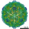
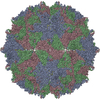
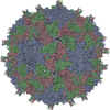
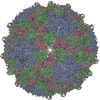
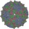
| Entry | Database: PDB / ID: 6eit | ||||||||||||
|---|---|---|---|---|---|---|---|---|---|---|---|---|---|
| Title | Coxsackievirus A24v in complex with the D1-D2 fragment of ICAM-1 | ||||||||||||
 Components Components |
| ||||||||||||
 Keywords Keywords | VIRUS / Enterovirus / Receptor / Complex / picornavirus | ||||||||||||
| Function / homology |  Function and homology information Function and homology informationregulation of leukocyte mediated cytotoxicity / positive regulation of cellular extravasation / membrane to membrane docking / T cell activation via T cell receptor contact with antigen bound to MHC molecule on antigen presenting cell / leukocyte adhesion to vascular endothelial cell / adhesion of symbiont to host / cell-cell adhesion mediated by integrin / establishment of endothelial barrier / heterophilic cell-cell adhesion / leukocyte migration ...regulation of leukocyte mediated cytotoxicity / positive regulation of cellular extravasation / membrane to membrane docking / T cell activation via T cell receptor contact with antigen bound to MHC molecule on antigen presenting cell / leukocyte adhesion to vascular endothelial cell / adhesion of symbiont to host / cell-cell adhesion mediated by integrin / establishment of endothelial barrier / heterophilic cell-cell adhesion / leukocyte migration / leukocyte cell-cell adhesion / Interleukin-10 signaling / symbiont-mediated suppression of host cytoplasmic pattern recognition receptor signaling pathway via inhibition of RIG-I activity / immunological synapse / negative regulation of endothelial cell apoptotic process / negative regulation of extrinsic apoptotic signaling pathway via death domain receptors / Integrin cell surface interactions / picornain 2A / symbiont-mediated suppression of host mRNA export from nucleus / symbiont genome entry into host cell via pore formation in plasma membrane / picornain 3C / T=pseudo3 icosahedral viral capsid / host cell cytoplasmic vesicle membrane / integrin binding / cellular response to amyloid-beta / Interferon gamma signaling / Immunoregulatory interactions between a Lymphoid and a non-Lymphoid cell / viral capsid / ribonucleoside triphosphate phosphatase activity / transmembrane signaling receptor activity / host cell / nucleoside-triphosphate phosphatase / extracellular matrix / virus receptor activity / channel activity / signaling receptor activity / Interleukin-4 and Interleukin-13 signaling / monoatomic ion transmembrane transport / positive regulation of ERK1 and ERK2 cascade / receptor-mediated virion attachment to host cell / cell adhesion / RNA helicase activity / endocytosis involved in viral entry into host cell / membrane raft / receptor ligand activity / external side of plasma membrane / symbiont-mediated activation of host autophagy / RNA-directed RNA polymerase / cysteine-type endopeptidase activity / focal adhesion / viral RNA genome replication / RNA-directed RNA polymerase activity / symbiont entry into host cell / virion attachment to host cell / host cell nucleus / structural molecule activity / cell surface / DNA-templated transcription / proteolysis / : / RNA binding / extracellular exosome / zinc ion binding / ATP binding / membrane / plasma membrane Similarity search - Function | ||||||||||||
| Biological species |  Homo sapiens (human) Homo sapiens (human) Coxsackievirus A24 Coxsackievirus A24 | ||||||||||||
| Method | ELECTRON MICROSCOPY / single particle reconstruction / cryo EM / Resolution: 3.9 Å | ||||||||||||
 Authors Authors | Hurdiss, D.L. / Ranson, N.A. | ||||||||||||
| Funding support |  Netherlands, Netherlands,  Germany, Germany,  United Kingdom, 3items United Kingdom, 3items
| ||||||||||||
 Citation Citation | Journal: Proc Natl Acad Sci U S A / Year: 2018 Title: Role of enhanced receptor engagement in the evolution of a pandemic acute hemorrhagic conjunctivitis virus. Authors: Jim Baggen / Daniel L Hurdiss / Georg Zocher / Nitesh Mistry / Richard W Roberts / Jasper J Slager / Hongbo Guo / Arno L W van Vliet / Maryam Wahedi / Kimberley Benschop / Erwin Duizer / ...Authors: Jim Baggen / Daniel L Hurdiss / Georg Zocher / Nitesh Mistry / Richard W Roberts / Jasper J Slager / Hongbo Guo / Arno L W van Vliet / Maryam Wahedi / Kimberley Benschop / Erwin Duizer / Cornelis A M de Haan / Erik de Vries / José M Casasnovas / Raoul J de Groot / Niklas Arnberg / Thilo Stehle / Neil A Ranson / Hendrik Jan Thibaut / Frank J M van Kuppeveld /   Abstract: Acute hemorrhagic conjunctivitis (AHC) is a painful, contagious eye disease, with millions of cases in the last decades. Coxsackievirus A24 (CV-A24) was not originally associated with human disease, ...Acute hemorrhagic conjunctivitis (AHC) is a painful, contagious eye disease, with millions of cases in the last decades. Coxsackievirus A24 (CV-A24) was not originally associated with human disease, but in 1970 a pathogenic "variant" (CV-A24v) emerged, which is now the main cause of AHC. Initially, this variant circulated only in Southeast Asia, but it later spread worldwide, accounting for numerous AHC outbreaks and two pandemics. While both CV-A24 variant and nonvariant strains still circulate in humans, only variant strains cause AHC for reasons that are yet unknown. Since receptors are important determinants of viral tropism, we set out to map the CV-A24 receptor repertoire and establish whether changes in receptor preference have led to the increased pathogenicity and rapid spread of CV-A24v. Here, we identify ICAM-1 as an essential receptor for both AHC-causing and non-AHC strains. We provide a high-resolution cryo-EM structure of a virus-ICAM-1 complex, which revealed critical ICAM-1-binding residues. These data could help identify a possible conserved mode of receptor engagement among ICAM-1-binding enteroviruses and rhinoviruses. Moreover, we identify a single capsid substitution that has been adopted by all pandemic CV-A24v strains and we reveal that this adaptation enhances the capacity of CV-A24v to bind sialic acid. Our data elucidate the CV-A24v receptor repertoire and point to a role of enhanced receptor engagement in the adaptation to the eye, possibly enabling pandemic spread. | ||||||||||||
| History |
|
- Structure visualization
| Movie |
Movie viewer |
|---|---|
| Structure viewer | Molecule: MolmilJmol/JSmol |
- Downloads & links
-Download
| PDBx/mmCIF format | 6eit.cif.gz | 162.4 KB | Display | PDBx/mmCIF format |
|---|---|---|---|---|
| PDB format | pdb6eit.ent.gz | 127.1 KB | Display | PDB format |
| PDBx/mmJSON format | 6eit.json.gz | Tree view | PDBx/mmJSON format | |
| Others |  Other downloads Other downloads |
-Validation report
| Arichive directory | https://data.pdbj.org/pub/pdb/validation_reports/ei/6eitftp://data.pdbj.org/pub/pdb/validation_reports/ei/6eit | HTTPS FTP |
|---|
-Related structure data
| Related structure data | 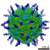 3880MC M: map data used to model this data C: citing same article ( |
|---|---|
| Similar structure data |
-Links
 PDBj
PDBj


- Assembly
| Deposited unit | 
|
|---|---|
| 1 | x 60
|
| Symmetry | Point symmetry: (Schoenflies symbol: I (icosahedral)) |
-Components
| #1: Protein | Mass: 34378.371 Da / Num. of mol.: 1 / Source method: isolated from a natural source / Source: (natural) Coxsackievirus A24 / Cell line: Normal human conjunctival (NHC) cells / References: UniProt: G3C8J7, UniProt: V9VEF3*PLUS |
|---|---|
| #2: Protein | Mass: 29817.412 Da / Num. of mol.: 1 / Source method: isolated from a natural source / Source: (natural) Coxsackievirus A24 / Cell line: Normal human conjunctival (NHC) cells / References: UniProt: A0A088F913, UniProt: V9VEF3*PLUS |
| #3: Protein | Mass: 26637.746 Da / Num. of mol.: 1 / Source method: isolated from a natural source / Source: (natural) Coxsackievirus A24 / Cell line: Normal human conjunctival (NHC) cells / References: UniProt: Q0GYP7, UniProt: V9VEF3*PLUS |
| #4: Protein | Mass: 9297.600 Da / Num. of mol.: 1 Source method: isolated from a genetically manipulated source Source: (gene. exp.) Homo sapiens (human) / Gene: ICAM1 / Cell line (production host): CHO Lec3.2.8.1 cells / Production host:   Cricetulus griseus (Chinese hamster) / References: UniProt: P05362 Cricetulus griseus (Chinese hamster) / References: UniProt: P05362 |
| Has protein modification | Y |
-Experimental details
-Experiment
| Experiment | Method: ELECTRON MICROSCOPY |
|---|---|
| EM experiment | Aggregation state: PARTICLE / 3D reconstruction method: single particle reconstruction |
- Sample preparation
| Component |
| ||||||||||||||||||||||||
|---|---|---|---|---|---|---|---|---|---|---|---|---|---|---|---|---|---|---|---|---|---|---|---|---|---|
| Molecular weight | Value: 8 MDa / Experimental value: NO | ||||||||||||||||||||||||
| Source (natural) |
| ||||||||||||||||||||||||
| Source (recombinant) | Organism: Cricetulus griseus (Chinese hamster) | ||||||||||||||||||||||||
| Details of virus | Empty: NO / Enveloped: NO / Isolate: STRAIN / Type: VIRION | ||||||||||||||||||||||||
| Natural host | Organism: Homo sapiens | ||||||||||||||||||||||||
| Virus shell | Diameter: 300 nm / Triangulation number (T number): 3 | ||||||||||||||||||||||||
| Buffer solution | pH: 7.5 Details: TBS buffer (Coxsackievirus A24v) Phosphate buffer (ICAM-1 D1-D2) | ||||||||||||||||||||||||
| Specimen | Conc.: 10 mg/ml / Embedding applied: NO / Shadowing applied: NO / Staining applied: NO / Vitrification applied: YES | ||||||||||||||||||||||||
| Specimen support | Grid material: COPPER / Grid mesh size: 400 divisions/in. Grid type: Lacey grids coated in a 3 nm carbon film (Agar Scientific, UK) | ||||||||||||||||||||||||
| Vitrification | Instrument: LEICA EM GP / Cryogen name: ETHANE / Humidity: 80 % / Chamber temperature: 8 K Details: On-grid binding of the receptor was performed by applying 3 microliters of ICAM-1 (9.85 mg/ml) to the pre-blotted, virus-containing grid, and leaving for 30 seconds before blotting and freezing |
- Electron microscopy imaging
| Experimental equipment |  Model: Titan Krios / Image courtesy: FEI Company |
|---|---|
| Microscopy | Model: FEI TITAN KRIOS |
| Electron gun | Electron source:  FIELD EMISSION GUN / Accelerating voltage: 300 kV / Illumination mode: FLOOD BEAM FIELD EMISSION GUN / Accelerating voltage: 300 kV / Illumination mode: FLOOD BEAM |
| Electron lens | Mode: BRIGHT FIELD |
| Specimen holder | Cryogen: NITROGEN / Specimen holder model: FEI TITAN KRIOS AUTOGRID HOLDER |
| Image recording | Average exposure time: 1.5 sec. / Electron dose: 60 e/Å2 / Detector mode: INTEGRATING / Film or detector model: FEI FALCON III (4k x 4k) / Num. of grids imaged: 1 / Num. of real images: 2652 |
- Processing
| EM software | Name: RELION / Version: 2 / Category: 3D reconstruction |
|---|---|
| CTF correction | Details: gCTF / Type: PHASE FLIPPING AND AMPLITUDE CORRECTION |
| Symmetry | Point symmetry: I (icosahedral) |
| 3D reconstruction | Resolution: 3.9 Å / Resolution method: FSC 0.143 CUT-OFF / Num. of particles: 26311 / Symmetry type: POINT |
| Atomic model building | Protocol: OTHER / Space: REAL |
