[English] 日本語
Yorodumi
- PDB-6egn: Crystal Structure of a Three-stranded Coiled Coil Peptide Contain... -

+
Open data


ID or keywords:

Loading...

-
Basic information

Entry
Database: PDB / ID: 6egn
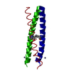
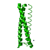
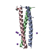
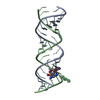
TitleCrystal Structure of a Three-stranded Coiled Coil Peptide Containing a Trigonal Planar Hg(II)S3 Site Modified by D-Leu in the Second Coordination Sphere
ComponentsHg(II)(GRAND CoilSerL16CL19(DLE))3-
KeywordsDE NOVO PROTEIN / De Novo Three-stranded Helical Coiled Coil Peptide / Hg(II)S3 complex / D-amino acids / D-Leu / Trigonal Planar Hg(II) Structure
Function / homology: / PROLINE
Function and homology information
Biological speciessynthetic construct (others)
MethodX-RAY DIFFRACTION / SYNCHROTRON / Resolution: 1.84 Å
AuthorsRuckthong, L. / Stuckey, J.A. / Pecoraro, V.L.
Funding support United States, 1items
OrganizationGrant numberCountry
National Institutes of Health/National Human Genome Research Institute (NIH/NHGRI)R01ES012236 United States
CitationJournal: Chemistry / Year: 2019
Title: How Outer Coordination Sphere Modifications Can Impact Metal Structures in Proteins: A Crystallographic Evaluation.
Authors: Ruckthong, L. / Stuckey, J.A. / Pecoraro, V.L.
History
DepositionAug 20, 2018Deposition site: RCSB / Processing site: RCSB
Revision 1.0Apr 3, 2019Provider: repository / Type: Initial release
Revision 1.1May 8, 2019Group: Data collection / Database references / Category: citation / citation_author
Item: _citation.journal_id_ISSN / _citation.title ..._citation.journal_id_ISSN / _citation.title / _citation_author.identifier_ORCID / _citation_author.name
Revision 1.2May 22, 2019Group: Data collection / Database references / Category: citation / citation_author
Item: _citation.journal_volume / _citation.page_first ..._citation.journal_volume / _citation.page_first / _citation.page_last / _citation_author.identifier_ORCID
Revision 1.3Dec 18, 2019Group: Author supporting evidence / Category: pdbx_audit_support / Item: _pdbx_audit_support.funding_organization

-
Structure visualization

Structure viewerMolecule:
MolmilJmol/JSmol

Downloads & links

-
Assembly

Deposited unit
A: Hg(II)(GRAND CoilSerL16CL19(DLE))3-
B: Hg(II)(GRAND CoilSerL16CL19(DLE))3-
C: Hg(II)(GRAND CoilSerL16CL19(DLE))3-
hetero molecules


Theoretical massNumber of molelcules
Total (without water)16,13912
Polymers12,4173
Non-polymers3,7229
Water2,972165
1


  • Idetical with deposited unit
  • defined by author&software
  • Evidence: mass spectrometry
TypeNameSymmetry operationNumber
identity operation1_555x,y,z1
Buried area5660 Å2
ΔGint-144 kcal/mol
Surface area8020 Å2
MethodPISA
Unit cell
Length a, b, c (Å)32.636, 80.508, 88.730
Angle α, β, γ (deg.)90.000, 90.000, 90.000
Int Tables number19
Space group name H-MP212121

-
Components

-
Protein/peptide , 1 types, 3 molecules ABC

#1: Protein/peptide Hg(II)(GRAND CoilSerL16CL19(DLE))3-


Mass: 4138.846 Da / Num. of mol.: 3 / Source method: obtained synthetically / Source: (synth.) synthetic construct (others)

-
Non-polymers , 6 types, 174 molecules

#2: Chemical ChemComp-ZN / ZINC ION


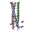
Mass: 65.409 Da / Num. of mol.: 3 / Source method: obtained synthetically / Formula: Zn
#3: Chemical ChemComp-PRO / PROLINE / Proline


Type: L-peptide linking / Mass: 115.130 Da / Num. of mol.: 2 / Source method: obtained synthetically / Formula: C5H9NO2
#4: Chemical ChemComp-15P / POLYETHYLENE GLYCOL (N=34) / PEG 1500 / Polyethylene glycol


Mass: 1529.829 Da / Num. of mol.: 2 / Source method: obtained synthetically / Formula: C69H140O35 / Comment: precipitant*YM
#5: Chemical ChemComp-CL / CHLORIDE ION / Chloride


Mass: 35.453 Da / Num. of mol.: 1 / Source method: obtained synthetically / Formula: Cl
#6: Chemical ChemComp-HG / MERCURY (II) ION / Mercury (element)


Mass: 200.590 Da / Num. of mol.: 1 / Source method: obtained synthetically / Formula: Hg
#7: Water ChemComp-HOH / water / Water


Mass: 18.015 Da / Num. of mol.: 165 / Source method: isolated from a natural source / Formula: H2O

-
Experimental details

-
Experiment

ExperimentMethod: X-RAY DIFFRACTION / Number of used crystals: 1

-
Sample preparation

CrystalDensity Matthews: 4.69 Å3/Da / Density % sol: 73.8 %
Crystal growTemperature: 293 K / Method: vapor diffusion, sitting drop
Details: 0.2 M L-Proline, 0.1 M HEPES, pH 7.5, 10% w/v PEG3350
PH range: 7.5-8.5

-
Data collection

DiffractionMean temperature: 100 K
Diffraction sourceSource: SYNCHROTRON / Site: APS / Beamline: 21-ID-G / Wavelength: 0.97856 Å
DetectorType: MARMOSAIC 300 mm CCD / Detector: CCD / Date: Nov 19, 2014
RadiationMonochromator: diamond(111) / Protocol: SINGLE WAVELENGTH / Monochromatic (M) / Laue (L): M / Scattering type: x-ray
Radiation wavelengthWavelength: 0.97856 Å / Relative weight: 1
ReflectionResolution: 1.84→50 Å / Num. obs: 20279 / % possible obs: 97 % / Redundancy: 8.8 % / Biso Wilson estimate: 36.53 Å2 / Rmerge(I) obs: 0.102 / Rpim(I) all: 0.04 / Rrim(I) all: 0.11 / Χ2: 0.96 / Net I/σ(I): 6.7 / Num. measured all: 178615
Reflection shell

Diffraction-ID: 1

Resolution (Å)Redundancy (%)Rmerge(I) obsNum. unique obsCC1/2Rpim(I) allRrim(I) allΧ2% possible all
1.84-1.878.20.46810110.9050.1740.5010.63799.4
1.87-1.9190.42310350.9270.1520.4510.69699.6
1.91-1.949.30.379910.9340.1320.3940.74199.5
1.94-1.989.50.32910090.9640.1160.350.72299.4
1.98-2.039.50.28910650.9550.1010.3070.78199.4
2.03-2.079.20.2499950.9680.0890.2650.84199.1
2.07-2.129.40.21510070.9620.0770.2290.89199.2
2.12-2.189.30.19410330.9780.070.2070.91898.8
2.18-2.259.20.1710030.9750.0620.1820.94898.6
2.25-2.329.20.15510230.9770.0580.1660.9698.1
2.32-2.48.90.1469930.9790.0550.1571.05197.7
2.4-2.590.13910390.9860.0520.1491.06698.1
2.5-2.618.80.1299880.9840.0490.1381.10197
2.61-2.758.90.12210190.9890.0460.1311.13897
2.75-2.928.90.1169990.9830.0450.1251.13896.1
2.92-3.158.60.10810130.9850.0410.1171.18195.7
3.15-3.468.50.09510060.9890.0360.1021.14395.1
3.46-3.9680.08910150.9920.0340.0961.14395
3.96-4.997.80.08610210.990.0340.0931.09893.4
4.99-507.10.08710140.9870.0370.0951.14986.3

-
Processing

Software
NameVersionClassification
HKL-2000data scaling
BUSTER-TNT2.10.2refinement
PDB_EXTRACT3.24data extraction
HKL-2000data reduction
AutoSolphasing
HKL-2000data collection
RefinementResolution: 1.84→19.14 Å / Cor.coef. Fo:Fc: 0.946 / Cor.coef. Fo:Fc free: 0.949 / Rfactor Rfree error: 0 / SU R Cruickshank DPI: 0.097 / Cross valid method: THROUGHOUT / σ(F): 0 / SU R Blow DPI: 0.109 / SU Rfree Blow DPI: 0.099 / SU Rfree Cruickshank DPI: 0.091
RfactorNum. reflection% reflectionSelection details
Rfree0.209 980 4.85 %RANDOM
Rwork0.197 ---
obs0.198 20219 96.2 %-
Displacement parametersBiso max: 147.85 Å2 / Biso mean: 46.02 Å2 / Biso min: 22.53 Å2
Baniso -1Baniso -2Baniso -3
1--7.19 Å20 Å20 Å2
2---1.391 Å20 Å2
3---8.5809 Å2
Refine analyzeLuzzati coordinate error obs: 0.24 Å
Refinement stepCycle: final / Resolution: 1.84→19.14 Å
ProteinNucleic acidLigandSolventTotal
Num. atoms862 0 38 165 1065
Biso mean--95.83 54.75 -
Num. residues----114
Refine LS restraints
Refine-IDTypeNumberRestraint functionWeightDev ideal
X-RAY DIFFRACTIONt_dihedral_angle_d388SINUSOIDAL2
X-RAY DIFFRACTIONt_trig_c_planes31HARMONIC2
X-RAY DIFFRACTIONt_gen_planes131HARMONIC5
X-RAY DIFFRACTIONt_it955HARMONIC20
X-RAY DIFFRACTIONt_nbd2SEMIHARMONIC5
X-RAY DIFFRACTIONt_improper_torsion
X-RAY DIFFRACTIONt_pseud_angle
X-RAY DIFFRACTIONt_chiral_improper_torsion119SEMIHARMONIC5
X-RAY DIFFRACTIONt_sum_occupancies
X-RAY DIFFRACTIONt_utility_distance
X-RAY DIFFRACTIONt_utility_angle
X-RAY DIFFRACTIONt_utility_torsion
X-RAY DIFFRACTIONt_ideal_dist_contact1184SEMIHARMONIC4
X-RAY DIFFRACTIONt_bond_d967HARMONIC20.01
X-RAY DIFFRACTIONt_angle_deg1291HARMONIC21.01
X-RAY DIFFRACTIONt_omega_torsion2.25
X-RAY DIFFRACTIONt_other_torsion18.52
LS refinement shellResolution: 1.84→1.94 Å / Rfactor Rfree error: 0 / Total num. of bins used: 10
RfactorNum. reflection% reflection
Rfree0.274 127 4.52 %
Rwork0.227 2682 -
all0.229 2809 -
obs--93.29 %
Refinement TLS params.

L11: 0 °2 / Method: refined / Refine-ID: X-RAY DIFFRACTION

IDL122)L132)L222)L232)L332)S11 (Å °)S12 (Å °)S13 (Å °)S21 (Å °)S22 (Å °)S23 (Å °)S31 (Å °)S32 (Å °)S33 (Å °)T112)T122)T132)T222)T232)T332)Origin x (Å)Origin y (Å)Origin z (Å)
11.0074-1.4675.1197-4.79364.2396-0.0044-0.2824-0.14540.2124-0.5755-0.6338-0.07280.50350.5798-0.0708-0.0621-0.0630.04710.12550.03152.50265.292417.9375
20.1323-0.56552.8621-3.84534.5143-0.02450.0176-0.08760.15740.0476-0.0275-0.0799-0.1445-0.023-0.0003-0.0238-0.0281-0.0050.0476-0.0074-6.89845.240215.1706
31.2519-1.27041.0749-2.13543.4425-0.1215-0.0293-0.1765-0.3627-0.103-0.22950.6890.08650.2245-0.01950.03760.0557-0.00950.05290.0502-1.35630.015210.7418
Refinement TLS group
IDRefine-IDRefine TLS-IDSelection detailsAuth asym-IDAuth seq-ID
1X-RAY DIFFRACTION1{A|*}A0 - 37
2X-RAY DIFFRACTION2{B|*}B0 - 37
3X-RAY DIFFRACTION3{C|*}C0 - 37

+
About Yorodumi

-
News

-
Feb 9, 2022. New format data for meta-information of EMDB entries

New format data for meta-information of EMDB entries

  • Version 3 of the EMDB header file is now the official format.
  • The previous official version 1.9 will be removed from the archive.

Related info.:EMDB header

External links:wwPDB to switch to version 3 of the EMDB data model

-
Aug 12, 2020. Covid-19 info

Covid-19 info

URL: https://pdbj.org/emnavi/covid19.php

New page: Covid-19 featured information page in EM Navigator.

Related info.:Covid-19 info / Mar 5, 2020. Novel coronavirus structure data

+
Mar 5, 2020. Novel coronavirus structure data

Novel coronavirus structure data

Related info.:Yorodumi Speices / Aug 12, 2020. Covid-19 info

External links:COVID-19 featured content - PDBj / Molecule of the Month (242):Coronavirus Proteases

+
Jan 31, 2019. EMDB accession codes are about to change! (news from PDBe EMDB page)

EMDB accession codes are about to change! (news from PDBe EMDB page)

  • The allocation of 4 digits for EMDB accession codes will soon come to an end. Whilst these codes will remain in use, new EMDB accession codes will include an additional digit and will expand incrementally as the available range of codes is exhausted. The current 4-digit format prefixed with “EMD-” (i.e. EMD-XXXX) will advance to a 5-digit format (i.e. EMD-XXXXX), and so on. It is currently estimated that the 4-digit codes will be depleted around Spring 2019, at which point the 5-digit format will come into force.
  • The EM Navigator/Yorodumi systems omit the EMD- prefix.

Related info.:Q: What is EMD? / ID/Accession-code notation in Yorodumi/EM Navigator

External links:EMDB Accession Codes are Changing Soon! / Contact to PDBj

+
Jul 12, 2017. Major update of PDB

Major update of PDB

  • wwPDB released updated PDB data conforming to the new PDBx/mmCIF dictionary.
  • This is a major update changing the version number from 4 to 5, and with Remediation, in which all the entries are updated.
  • In this update, many items about electron microscopy experimental information are reorganized (e.g. em_software).
  • Now, EM Navigator and Yorodumi are based on the updated data.

External links:wwPDB Remediation / Enriched Model Files Conforming to OneDep Data Standards Now Available in the PDB FTP Archive

-
Yorodumi

Thousand views of thousand structures

  • Yorodumi is a browser for structure data from EMDB, PDB, SASBDB, etc.
  • This page is also the successor to EM Navigator detail page, and also detail information page/front-end page for Omokage search.
  • The word "yorodu" (or yorozu) is an old Japanese word meaning "ten thousand". "mi" (miru) is to see.

Related info.:EMDB / PDB / SASBDB / Comparison of 3 databanks / Yorodumi Search / Aug 31, 2016. New EM Navigator & Yorodumi / Yorodumi Papers / Jmol/JSmol / Function and homology information / Changes in new EM Navigator and Yorodumi

Read more