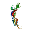
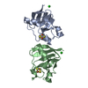
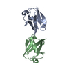
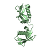
LYSINE-SPECIFICDEMETHYLASE2A / CXXC-TYPE ZINC FINGER PROTEIN 8 / F-BOX AND LEUCINE-RICH REPEAT PROTEIN 11 / F-BOX PROTEIN FBL7 / F- ...CXXC-TYPE ZINC FINGER PROTEIN 8 / F-BOX AND LEUCINE-RICH REPEAT PROTEIN 11 / F-BOX PROTEIN FBL7 / F-BOX PROTEIN LILINA / F-BOX/LRR-REPEAT PROTEIN 11 / JMJC DOMAIN-CONTAINING HISTONE DEMETHYLATION PROTEIN 1A / [HISTONE-H3]-LYSINE-36 DEMETHYLASE 1A
Mass: 13319.508 Da / Num. of mol.: 2 / Fragment: CXXC AND PHD DOMAIN, RESIDUES 567-681 Source method: isolated from a genetically manipulated source Source: (gene. exp.) HOMO SAPIENS (human) / Plasmid: PNIC28-BSA4 / Production host: ESCHERICHIA COLI (E. coli) / Strain (production host): BL21(DE3) / Variant (production host): R3-PRARE2 References: UniProt: Q9Y2K7, [histone H3]-dimethyl-L-lysine36 demethylase
Monochromator: DOUBLE CRYSTAL / Protocol: SINGLE WAVELENGTH / Monochromatic (M) / Laue (L): M / Scattering type: x-ray
Radiation wavelength
Wavelength: 0.92 Å / Relative weight: 1
Reflection
Resolution: 2.24→90.79 Å / Num. obs: 13091 / % possible obs: 98.7 % / Redundancy: 7.5 % / Biso Wilson estimate: 39.61 Å2 / Rmerge(I) obs: 0.08 / Net I/σ(I): 16
Reflection shell
Resolution: 2.24→2.36 Å / Redundancy: 7.5 % / Rmerge(I) obs: 0.49 / Mean I/σ(I) obs: 3.8 / % possible all: 98.2
-
Processing
Software
Name
Version
Classification
BUSTER
2.11.2
refinement
autoPROC
datareduction
AP_SCALE
datascaling
SHARP
phasing
Refinement
Method to determine structure: SAD Starting model: NONE Resolution: 2.24→90.79 Å / Cor.coef. Fo:Fc: 0.9194 / Cor.coef. Fo:Fc free: 0.8894 / SU R Cruickshank DPI: 0.225 / Cross valid method: THROUGHOUT / σ(F): 0 / SU R Blow DPI: 0.246 / SU Rfree Blow DPI: 0.182 / SU Rfree Cruickshank DPI: 0.176 Details: IDEAL-DIST CONTACT TERM CONTACT SETUP. RESIDUE TYPES WITHOUT CCP4 ATOM TYPE IN LIBRARY=ZN. NUMBER WITH APPROX DEFAULT CCP4 ATOM TYPE=0. NUMBER TREATED BY BAD NON-BONDED CONTACTS=8.
Rfactor
Num. reflection
% reflection
Selection details
Rfree
0.2209
643
4.93 %
RANDOM
Rwork
0.1969
-
-
-
obs
0.1981
13045
98.98 %
-
Displacement parameters
Biso mean: 48.63 Å2
Baniso -1
Baniso -2
Baniso -3
1-
-11.9938 Å2
0 Å2
6.3255 Å2
2-
-
8.5589 Å2
0 Å2
3-
-
-
3.4349 Å2
Refine analyze
Luzzati coordinate error obs: 0.311 Å
Refinement step
Cycle: LAST / Resolution: 2.24→90.79 Å
Protein
Nucleic acid
Ligand
Solvent
Total
Num. atoms
1564
0
12
106
1682
Refine LS restraints
Refine-ID
Type
Dev ideal
Number
Restraint function
Weight
X-RAY DIFFRACTION
t_bond_d
0.01
1592
HARMONIC
2
X-RAY DIFFRACTION
t_angle_deg
1.09
2139
HARMONIC
2
X-RAY DIFFRACTION
t_dihedral_angle_d
750
SINUSOIDAL
2
X-RAY DIFFRACTION
t_incorr_chiral_ct
X-RAY DIFFRACTION
t_pseud_angle
X-RAY DIFFRACTION
t_trig_c_planes
32
HARMONIC
2
X-RAY DIFFRACTION
t_gen_planes
238
HARMONIC
5
X-RAY DIFFRACTION
t_it
1592
HARMONIC
20
X-RAY DIFFRACTION
t_nbd
2
SEMIHARMONIC
5
X-RAY DIFFRACTION
t_omega_torsion
3.41
X-RAY DIFFRACTION
t_other_torsion
2.95
X-RAY DIFFRACTION
t_improper_torsion
X-RAY DIFFRACTION
t_chiral_improper_torsion
204
SEMIHARMONIC
5
X-RAY DIFFRACTION
t_sum_occupancies
X-RAY DIFFRACTION
t_utility_distance
X-RAY DIFFRACTION
t_utility_angle
X-RAY DIFFRACTION
t_utility_torsion
X-RAY DIFFRACTION
t_ideal_dist_contact
1756
SEMIHARMONIC
4
LS refinement shell
Resolution: 2.24→2.42 Å / Total num. of bins used: 7
Rfactor
Num. reflection
% reflection
Rfree
0.2343
136
5.1 %
Rwork
0.1996
2530
-
all
0.2015
2666
-
obs
-
-
98.98 %
Refinement TLS params.
Method: refined / Refine-ID: X-RAY DIFFRACTION
ID
L11 (°2)
L12 (°2)
L13 (°2)
L22 (°2)
L23 (°2)
L33 (°2)
S11 (Å °)
S12 (Å °)
S13 (Å °)
S21 (Å °)
S22 (Å °)
S23 (Å °)
S31 (Å °)
S32 (Å °)
S33 (Å °)
T11 (Å2)
T12 (Å2)
T13 (Å2)
T22 (Å2)
T23 (Å2)
T33 (Å2)
Origin x (Å)
Origin y (Å)
Origin z (Å)
1
0.3334
-0.6868
1.1002
2.0309
-2.9205
3.6596
0.0444
-0.0131
0.0308
0.205
-0.0626
-0.0736
-0.3318
0.2827
0.0181
0.0153
0.0121
-0.0536
-0.0778
-0.0313
-0.0405
4.191
17.106
65.8942
2
1.4599
1.5757
-2.1276
2.5425
-2.8803
4.3012
0.0013
0.3406
0.0871
-0.1763
0.1028
-0.1209
0.1941
-0.0664
-0.1041
-0.0532
0.0064
0.0381
-0.0139
0.0218
-0.0864
-21.5774
-15.47
71.4614
Refinement TLS group
ID
Refine-ID
Refine TLS-ID
Selection details
1
X-RAY DIFFRACTION
1
CHAINA
2
X-RAY DIFFRACTION
2
CHAINB
+
About Yorodumi
-
News
-
Feb 9, 2022. New format data for meta-information of EMDB entries
New format data for meta-information of EMDB entries
Version 3 of the EMDB header file is now the official format.
The previous official version 1.9 will be removed from the archive.
In the structure databanks used in Yorodumi, some data are registered as the other names, "COVID-19 virus" and "2019-nCoV". Here are the details of the virus and the list of structure data.
Jan 31, 2019. EMDB accession codes are about to change! (news from PDBe EMDB page)
EMDB accession codes are about to change! (news from PDBe EMDB page)
The allocation of 4 digits for EMDB accession codes will soon come to an end. Whilst these codes will remain in use, new EMDB accession codes will include an additional digit and will expand incrementally as the available range of codes is exhausted. The current 4-digit format prefixed with “EMD-” (i.e. EMD-XXXX) will advance to a 5-digit format (i.e. EMD-XXXXX), and so on. It is currently estimated that the 4-digit codes will be depleted around Spring 2019, at which point the 5-digit format will come into force.
The EM Navigator/Yorodumi systems omit the EMD- prefix.
Related info.:Q: What is EMD? / ID/Accession-code notation in Yorodumi/EM Navigator
Yorodumi is a browser for structure data from EMDB, PDB, SASBDB, etc.
This page is also the successor to EM Navigator detail page, and also detail information page/front-end page for Omokage search.
The word "yorodu" (or yorozu) is an old Japanese word meaning "ten thousand". "mi" (miru) is to see.
Related info.:EMDB / PDB / SASBDB / Comparison of 3 databanks / Yorodumi Search / Aug 31, 2016. New EM Navigator & Yorodumi / Yorodumi Papers / Jmol/JSmol / Function and homology information / Changes in new EM Navigator and Yorodumi
 Movie
Movie Controller

 Yorodumi
 Open data
 Basic information
 Components
 Keywords
 Function and homology information
 HOMO SAPIENS (human)
 X-RAY DIFFRACTION /
  Authors
 Citation
 Structure visualization
 Downloads & links
 Other downloads
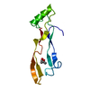


 PDBj
PDBj


 Assembly


 Mass: 62.068 Da / Num. of mol.: 1 / Source method: obtained synthetically / Formula: C2H6O2
 Mass: 65.409 Da / Num. of mol.: 8 / Source method: obtained synthetically / Formula: Zn
 Mass: 18.015 Da / Num. of mol.: 106 / Source method: isolated from a natural source / Formula: H2O
 Sample preparation
 / Beamline: I04-1 / Wavelength: 0.92
  Processing