 Movie
Movie Controller


+ Open data

- Basic information
| Entry | Database: PDB / ID: 6efg | ||||||
|---|---|---|---|---|---|---|---|
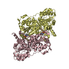
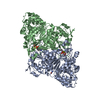
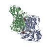
| Title | Pyruvate decarboxylase from Kluyveromyces lactis | ||||||
 Components Components | Pyruvate decarboxylase | ||||||
 Keywords Keywords | LYASE / pyruvate decarboxylase / thiamine diphosphate / substrate activation | ||||||
| Function / homology |  Function and homology information Function and homology informationpyruvate decarboxylase / : / pyruvate decarboxylase activity / thiamine pyrophosphate binding / magnesium ion binding / nucleus / cytosol Similarity search - Function | ||||||
| Biological species |  Kluyveromyces lactis (yeast) Kluyveromyces lactis (yeast) | ||||||
| Method |  X-RAY DIFFRACTION / SYNCHROTRON / MOLECULAR REPLACEMENT / Resolution: 2.04 Å X-RAY DIFFRACTION / SYNCHROTRON / MOLECULAR REPLACEMENT / Resolution: 2.04 Å | ||||||
 Authors Authors | Kutter, S. / Konig, S. | ||||||
 Citation Citation | Journal: To be Published Title: The crystal structures of pyruvate decarboxylase from Kluyveromyces lactis in the absence of ligands and in the presence of the substrate surrogate pyruvamide Authors: Kutter, S. / Konig, S. #1: Journal: FEBS J. / Year: 2006Title: The crystal structure of pyruvate decarboxylase from Kluyveromyces lactis. Implications for the substrate activation mechanism of this enzyme. Authors: Kutter, S. / Wille, G. / Relle, S. / Weiss, M.S. / Hubner, G. / Konig, S. | ||||||
| History |
|
- Structure visualization
| Structure viewer | Molecule: MolmilJmol/JSmol |
|---|
- Downloads & links
-Download
| PDBx/mmCIF format | 6efg.cif.gz | 421.3 KB | Display | PDBx/mmCIF format |
|---|---|---|---|---|
| PDB format | pdb6efg.ent.gz | 342 KB | Display | PDB format |
| PDBx/mmJSON format | 6efg.json.gz | Tree view | PDBx/mmJSON format | |
| Others |  Other downloads Other downloads |
-Validation report
| Arichive directory | https://data.pdbj.org/pub/pdb/validation_reports/ef/6efgftp://data.pdbj.org/pub/pdb/validation_reports/ef/6efg | HTTPS FTP |
|---|
-Related structure data
| Related structure data |  6efhC 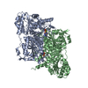 2vk4S S: Starting model for refinement C: citing same article ( |
|---|---|
| Similar structure data |

-Links
 PDBj
PDBj


- Assembly
| Deposited unit | 
| ||||||||
|---|---|---|---|---|---|---|---|---|---|
| 1 | 
| ||||||||
| 2 | 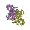
| ||||||||
| Unit cell |
|
-Components
| #1: Protein | Mass: 61716.762 Da / Num. of mol.: 4 / Source method: isolated from a natural source Source: (natural) Kluyveromyces lactis (strain ATCC 8585 / CBS 2359 / DSM 70799 / NBRC 1267 / NRRL Y-1140 / WM37) (yeast)Strain: ATCC 8585 / CBS 2359 / DSM 70799 / NBRC 1267 / NRRL Y-1140 / WM37 References: UniProt: Q12629, pyruvate decarboxylase #2: Chemical | ChemComp-TPP /   Mass: 425.314 Da / Num. of mol.: 4 / Source method: obtained synthetically / Formula: C12H19N4O7P2S Mass: 425.314 Da / Num. of mol.: 4 / Source method: obtained synthetically / Formula: C12H19N4O7P2S#3: Chemical | ChemComp-MG /   Mass: 24.305 Da / Num. of mol.: 4 / Source method: obtained synthetically / Formula: Mg Mass: 24.305 Da / Num. of mol.: 4 / Source method: obtained synthetically / Formula: Mg#4: Water | ChemComp-HOH / |  Mass: 18.015 Da / Num. of mol.: 308 / Source method: isolated from a natural source / Formula: H2O Mass: 18.015 Da / Num. of mol.: 308 / Source method: isolated from a natural source / Formula: H2O |
|---|
-Experimental details
-Experiment
| Experiment | Method: X-RAY DIFFRACTION / Number of used crystals: 1 |
|---|
- Sample preparation
| Crystal | Density Matthews: 2.37 Å3/Da / Density % sol: 48 % |
|---|---|
| Crystal grow | Temperature: 281.15 K / Method: vapor diffusion, hanging drop / pH: 6.25 Details: 2 mg/mL KlPDC in 2 mM MES, 18 mM citrate, pH 6.25, 4 mM thiamine diphosphate, 4 mM magnesium sulfate, 2 mM DTT, 1:1 with mother liquor (18-24% w/v PEG2000/PEG6000), ~25 days at 8 degrees C, ...Details: 2 mg/mL KlPDC in 2 mM MES, 18 mM citrate, pH 6.25, 4 mM thiamine diphosphate, 4 mM magnesium sulfate, 2 mM DTT, 1:1 with mother liquor (18-24% w/v PEG2000/PEG6000), ~25 days at 8 degrees C, soaked for 30 seconds in 2 uL mother liquor + 2 uL 15% w/v glycerol, 200 mM acetaldehyde prior to flash freezing. |
-Data collection
| Diffraction | Mean temperature: 100 K |
|---|---|
| Diffraction source | Source: SYNCHROTRON / Site: EMBL/DESY, HAMBURG  / Beamline: X12 / Wavelength: 1.00001 Å / Beamline: X12 / Wavelength: 1.00001 Å |
| Detector | Type: MARMOSAIC 225 mm CCD / Detector: CCD / Date: Nov 21, 2008 |
| Radiation | Monochromator: Si(111) / Protocol: SINGLE WAVELENGTH / Monochromatic (M) / Laue (L): M / Scattering type: x-ray |
| Radiation wavelength | Wavelength: 1.00001 Å / Relative weight: 1 |
| Reflection | Resolution: 2.04→38.25 Å / Num. obs: 126964 / % possible obs: 93.2 % / Redundancy: 2.7 % / Biso Wilson estimate: 24.75 Å2 / CC1/2: 0.996 / Rmerge(I) obs: 0.086 / Rpim(I) all: 0.065 / Rrim(I) all: 0.109 / Net I/σ(I): 8.6 |
| Reflection shell | Resolution: 2.04→2.08 Å / Redundancy: 2.3 % / Rmerge(I) obs: 0.659 / Mean I/σ(I) obs: 1.7 / Num. unique obs: 4511 / CC1/2: 0.689 / Rpim(I) all: 0.508 / Rrim(I) all: 0.837 / % possible all: 66.8 |
- Processing
| Software |
| ||||||||||||||||||||||||||||||||||||||||||||||||||||||||||||||||||||||||||||||||||||||||||||||||||||||||||||||||||||||||||||||||||||||||||||||||||||||||||||||||||||||||||||||||||||||
|---|---|---|---|---|---|---|---|---|---|---|---|---|---|---|---|---|---|---|---|---|---|---|---|---|---|---|---|---|---|---|---|---|---|---|---|---|---|---|---|---|---|---|---|---|---|---|---|---|---|---|---|---|---|---|---|---|---|---|---|---|---|---|---|---|---|---|---|---|---|---|---|---|---|---|---|---|---|---|---|---|---|---|---|---|---|---|---|---|---|---|---|---|---|---|---|---|---|---|---|---|---|---|---|---|---|---|---|---|---|---|---|---|---|---|---|---|---|---|---|---|---|---|---|---|---|---|---|---|---|---|---|---|---|---|---|---|---|---|---|---|---|---|---|---|---|---|---|---|---|---|---|---|---|---|---|---|---|---|---|---|---|---|---|---|---|---|---|---|---|---|---|---|---|---|---|---|---|---|---|---|---|---|---|
| Refinement | Method to determine structure: MOLECULAR REPLACEMENT Starting model: PDB entry 2VK4 Resolution: 2.04→38.25 Å / Cor.coef. Fo:Fc: 0.939 / Cor.coef. Fo:Fc free: 0.891 / SU B: 9.157 / SU ML: 0.23 / Cross valid method: THROUGHOUT / ESU R: 0.25 / ESU R Free: 0.223 / Details: HYDROGENS HAVE BEEN ADDED IN THE RIDING POSITIONS
| ||||||||||||||||||||||||||||||||||||||||||||||||||||||||||||||||||||||||||||||||||||||||||||||||||||||||||||||||||||||||||||||||||||||||||||||||||||||||||||||||||||||||||||||||||||||
| Solvent computation | Ion probe radii: 0.8 Å / Shrinkage radii: 0.8 Å / VDW probe radii: 1.2 Å | ||||||||||||||||||||||||||||||||||||||||||||||||||||||||||||||||||||||||||||||||||||||||||||||||||||||||||||||||||||||||||||||||||||||||||||||||||||||||||||||||||||||||||||||||||||||
| Displacement parameters | Biso mean: 37.997 Å2
| ||||||||||||||||||||||||||||||||||||||||||||||||||||||||||||||||||||||||||||||||||||||||||||||||||||||||||||||||||||||||||||||||||||||||||||||||||||||||||||||||||||||||||||||||||||||
| Refinement step | Cycle: 1 / Resolution: 2.04→38.25 Å
| ||||||||||||||||||||||||||||||||||||||||||||||||||||||||||||||||||||||||||||||||||||||||||||||||||||||||||||||||||||||||||||||||||||||||||||||||||||||||||||||||||||||||||||||||||||||
| Refine LS restraints |
|