 Movie
Movie Controller

[English] 日本語
 Yorodumi
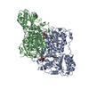
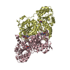
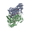
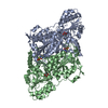
- PDB-1pvd: CRYSTAL STRUCTURE OF THE THIAMIN DIPHOSPHATE DEPENDENT ENZYME PYR... -
+ Open data

- Basic information
| Entry | Database: PDB / ID: 1pvd | ||||||
|---|---|---|---|---|---|---|---|
| Title | CRYSTAL STRUCTURE OF THE THIAMIN DIPHOSPHATE DEPENDENT ENZYME PYRUVATE DECARBOXYLASE FROM THE YEAST SACCHAROMYCES CEREVISIAE AT 2.3 ANGSTROMS RESOLUTION | ||||||
 Components Components | PYRUVATE DECARBOXYLASE | ||||||
 Keywords Keywords | LYASE (CARBON-CARBON) | ||||||
| Function / homology |  Function and homology information Function and homology informationphenylpyruvate decarboxylase / branched-chain-2-oxoacid decarboxylase / phenylpyruvate decarboxylase activity / branched-chain-2-oxoacid decarboxylase activity / indolepyruvate decarboxylase / pyruvate fermentation to ethanol / indolepyruvate decarboxylase activity / : / pyruvate decarboxylase activity / Lyases; Carbon-carbon lyases; Carboxy-lyases ...phenylpyruvate decarboxylase / branched-chain-2-oxoacid decarboxylase / phenylpyruvate decarboxylase activity / branched-chain-2-oxoacid decarboxylase activity / indolepyruvate decarboxylase / pyruvate fermentation to ethanol / indolepyruvate decarboxylase activity / : / pyruvate decarboxylase activity / Lyases; Carbon-carbon lyases; Carboxy-lyases / L-tryptophan catabolic process / branched-chain amino acid catabolic process / L-phenylalanine catabolic process / thiamine pyrophosphate binding / magnesium ion binding / nucleus / cytoplasm / cytosol Similarity search - Function | ||||||
| Biological species |  | ||||||
| Method |  X-RAY DIFFRACTION / Resolution: 2.3 Å X-RAY DIFFRACTION / Resolution: 2.3 Å | ||||||
 Authors Authors | Furey, W. / Arjunan, P. | ||||||
 Citation Citation | Journal: J.Mol.Biol. / Year: 1996 Title: Crystal structure of the thiamin diphosphate-dependent enzyme pyruvate decarboxylase from the yeast Saccharomyces cerevisiae at 2.3 A resolution. Authors: Arjunan, P. / Umland, T. / Dyda, F. / Swaminathan, S. / Furey, W. / Sax, M. / Farrenkopf, B. / Gao, Y. / Zhang, D. / Jordan, F. #1: Journal: Biochemistry and Physiology of Thiamin Diphosphate EnzymesYear: 1991 Title: Multiple Crystal Forms of Brewers' Yeast Pyruvate Decarboxylase: Characterization and Preliminary Crystallographic Analysis Authors: Dyda, F. / Furey, W. / Swaminathan, S. / Sax, M. / Farrenkopf, B. / Jordan, F. #2: Journal: Biochemistry and Physiology of Thiamin Diphosphate EnzymesYear: 1991 Title: Structure and Expression of Yeast Pyruvate Decarboxylase Structural Genes Authors: Hohmann, S. #3: Journal: J.Biol.Chem. / Year: 1990Title: Preliminary Crystallographic Data for the Thiamin Diphosphate-Dependent Enzyme Pyruvate Decarboxylase from Brewers' Yeast Authors: Dyda, F. / Furey, W. / Swaminathan, S. / Sax, M. / Farrenkopf, B. / Jordan, F. | ||||||
| History |
|
- Structure visualization
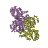
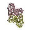
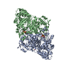
| Structure viewer | Molecule: MolmilJmol/JSmol |
|---|
- Downloads & links
-Download
| PDBx/mmCIF format | 1pvd.cif.gz | 228.9 KB | Display | PDBx/mmCIF format |
|---|---|---|---|---|
| PDB format | pdb1pvd.ent.gz | 179.1 KB | Display | PDB format |
| PDBx/mmJSON format | 1pvd.json.gz | Tree view | PDBx/mmJSON format | |
| Others |  Other downloads Other downloads |
-Validation report
| Arichive directory | https://data.pdbj.org/pub/pdb/validation_reports/pv/1pvdftp://data.pdbj.org/pub/pdb/validation_reports/pv/1pvd | HTTPS FTP |
|---|
-Related structure data
| Similar structure data |
|---|


-Links
 PDBj
PDBj


- Assembly
| Deposited unit | 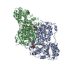
| ||||||||
|---|---|---|---|---|---|---|---|---|---|
| 1 |
| ||||||||
| Unit cell |
| ||||||||
| Noncrystallographic symmetry (NCS) | NCS oper: (Code: given Matrix: (-0.9115, -0.0493, -0.4084), Vector: Details | THE UNIT CELL CONTAINS TWO TETRAMERS, WITH ONE DIMER IN THE ASYMMETRIC UNIT. IN TERMS OF THE CRYSTAL AXES, THE TWO MONOMERS IN THE ASYMMETRIC UNIT (A AND B) ARE RELATED BY A NONCRYSTALLOGRAPHIC TWO-FOLD AXIS INCLINED BY 83.31 DEGREES TO THE B AXIS, AND WHOSE PROJECTION ON THE AC PLANE MAKES ANGLES OF 102.16 DEGREES WITH THE A AXIS, AND 14.23 DEGREES WITH THE C AXIS. THE NONCRYSTALLOGRAPHIC TWO-FOLD AXIS DOES NOT INTERSECT THE B AXIS, AS ITS CLOSEST APPROACH IS OFFSET BY 1.352 ANGSTROMS. THE TETRAMER THUS HAS ONLY APPROXIMATE 222 SYMMETRY, AS IT MUST INTERSECT THE B AXIS AND BE INCLINED TO IT BY 90 DEGREES FOR EXACT 222 SYMMETRY. MTRIX THE TRANSFORMATIONS PRESENTED ON MTRIX RECORDS BELOW DESCRIBE NON-CRYSTALLOGRAPHIC RELATIONSHIPS AMONG THE VARIOUS DOMAINS IN THIS ENTRY. APPLYING THE APPROPRIATE MTRIX TRANSFORMATION TO THE RESIDUES LISTED FIRST WILL YIELD APPROXIMATE COORDINATES FOR THE RESIDUES LISTED SECOND. APPLIED TO TRANSFORMED TO MTRIX RESIDUES RESIDUES RMSD M1 A 2 .. 556 B 2 .. 556 1.282 | |
-Components
| #1: Protein | Mass: 60741.148 Da / Num. of mol.: 2 Source method: isolated from a genetically manipulated source Source: (gene. exp.) References: UniProt: P06169, pyruvate decarboxylase #2: Chemical |   Mass: 24.305 Da / Num. of mol.: 2 / Source method: obtained synthetically / Formula: Mg Mass: 24.305 Da / Num. of mol.: 2 / Source method: obtained synthetically / Formula: Mg#3: Chemical |   Mass: 425.314 Da / Num. of mol.: 2 / Source method: obtained synthetically / Formula: C12H19N4O7P2S Mass: 425.314 Da / Num. of mol.: 2 / Source method: obtained synthetically / Formula: C12H19N4O7P2S#4: Water | ChemComp-HOH / |  Mass: 18.015 Da / Num. of mol.: 439 / Source method: isolated from a natural source / Formula: H2O Mass: 18.015 Da / Num. of mol.: 439 / Source method: isolated from a natural source / Formula: H2OCompound details | THE CATALYTIC CENTERS ARE LOCATED AT THE INTERFACE BETWEEN MONOMERS WITHIN EACH DIMER, WITH TWO ...THE CATALYTIC CENTERS ARE LOCATED AT THE INTERFACE BETWEEN MONOMERS WITHIN EACH DIMER, WITH TWO SITES PER DIMER AND FOUR PER TETRAMER. | |
|---|
-Experimental details
-Experiment
| Experiment | Method: X-RAY DIFFRACTION |
|---|
- Sample preparation
| Crystal | Density Matthews: 2.35 Å3/Da / Density % sol: 47.67 % | ||||||||||||||||||||||||||||||||||||||||||||||||
|---|---|---|---|---|---|---|---|---|---|---|---|---|---|---|---|---|---|---|---|---|---|---|---|---|---|---|---|---|---|---|---|---|---|---|---|---|---|---|---|---|---|---|---|---|---|---|---|---|---|
| Crystal grow | *PLUS Temperature: 4 ℃ / pH: 5.3 / Method: vapor diffusion, sitting drop | ||||||||||||||||||||||||||||||||||||||||||||||||
| Components of the solutions | *PLUS
|
-Data collection
| Reflection | Resolution: 2.3→40 Å / Num. obs: 46787 / % possible obs: 92.9 % / Observed criterion σ(F): 2 |
|---|---|
| Reflection | *PLUS % possible obs: 93 % / Num. measured all: 199754 / Rmerge(I) obs: 0.08 |
- Processing
| Software |
| ||||||||||||||||||||||||||||||||||||||||||||||||||||||||||||
|---|---|---|---|---|---|---|---|---|---|---|---|---|---|---|---|---|---|---|---|---|---|---|---|---|---|---|---|---|---|---|---|---|---|---|---|---|---|---|---|---|---|---|---|---|---|---|---|---|---|---|---|---|---|---|---|---|---|---|---|---|---|
| Refinement | Resolution: 2.3→10 Å / σ(F): 2 Details: REFERENCE 1 DESCRIBES THE TRANSFORMATION TO THE MONOCLINIC FORM AND ROTATION FUNCTION RESULTS. (APPROPRIATE FOR BOTH SACCHAROMYCES UVARUM AND SACCHAROMYCES CEREVISIAE). REFERENCE 3 DESCRIBES ...Details: REFERENCE 1 DESCRIBES THE TRANSFORMATION TO THE MONOCLINIC FORM AND ROTATION FUNCTION RESULTS. (APPROPRIATE FOR BOTH SACCHAROMYCES UVARUM AND SACCHAROMYCES CEREVISIAE). REFERENCE 3 DESCRIBES THE TRICLINIC CRYSTALS OF THE RELATED SACCHAROMYCES UVARUM FORM.
| ||||||||||||||||||||||||||||||||||||||||||||||||||||||||||||
| Refinement step | Cycle: LAST / Resolution: 2.3→10 Å
| ||||||||||||||||||||||||||||||||||||||||||||||||||||||||||||
| Refine LS restraints |
| ||||||||||||||||||||||||||||||||||||||||||||||||||||||||||||
| Refinement | *PLUS Num. reflection obs: 46787 | ||||||||||||||||||||||||||||||||||||||||||||||||||||||||||||
| Solvent computation | *PLUS | ||||||||||||||||||||||||||||||||||||||||||||||||||||||||||||
| Displacement parameters | *PLUS |
