 Movie
Movie Controller

[English] 日本語
 Yorodumi
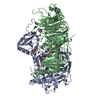
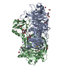
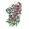
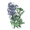
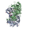
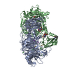
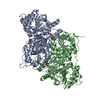
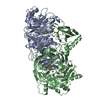
- PDB-2vk4: Crystal structure of pyruvate decarboxylase from Kluyveromyces lactis -
+ Open data

- Basic information
| Entry | Database: PDB / ID: 2vk4 | |||||||||
|---|---|---|---|---|---|---|---|---|---|---|
| Title | Crystal structure of pyruvate decarboxylase from Kluyveromyces lactis | |||||||||
 Components Components | PYRUVATE DECARBOXYLASE | |||||||||
 Keywords Keywords | LYASE / METAL-BINDING / DECARBOXYLASE / DIMER OF DIMERS / SUBSTRATE ACTIVATION / THIAMINE DIPHOSPHATE / TDP / TPP / MAGNESIUM / FLAVOPROTEIN / THIAMINE PYROPHOSPHATE / ASYMMETRIC ACTIVE SITES | |||||||||
| Function / homology |  Function and homology information Function and homology informationpyruvate decarboxylase / : / pyruvate decarboxylase activity / thiamine pyrophosphate binding / magnesium ion binding / nucleus / cytosol Similarity search - Function | |||||||||
| Biological species |  KLUYVEROMYCES LACTIS (yeast) KLUYVEROMYCES LACTIS (yeast) | |||||||||
| Method |  X-RAY DIFFRACTION / SYNCHROTRON / MOLECULAR REPLACEMENT / Resolution: 1.95 Å X-RAY DIFFRACTION / SYNCHROTRON / MOLECULAR REPLACEMENT / Resolution: 1.95 Å | |||||||||
 Authors Authors | Kutter, S. / Relle, S. / Wille, G. / Weiss, M.S. / Konig, S. | |||||||||
 Citation Citation | Journal: J.Mol.Catal., B Enzym. / Year: 2014 Title: Allosteric Activation of Pyruvate Decarboxylases. A Never-Ending Story. Authors: Konig, S. / Spinka, M. / Kutter, S. #1: Journal: FEBS J. / Year: 2006 Title: The Crystal Structure of Pyruvate Decarboxylase from Kluyveromyces Lactis. Implications for the Substrate Activation Mechanism of This Enzyme. Authors: Kutter, S. / Wille, G. / Relle, S. / Weiss, M.S. / Hubner, G. / Konig, S. | |||||||||
| History |
|
- Structure visualization
| Structure viewer | Molecule: MolmilJmol/JSmol |
|---|
- Downloads & links
-Download
| PDBx/mmCIF format | 2vk4.cif.gz | 458.6 KB | Display | PDBx/mmCIF format |
|---|---|---|---|---|
| PDB format | pdb2vk4.ent.gz | 372.2 KB | Display | PDB format |
| PDBx/mmJSON format | 2vk4.json.gz | Tree view | PDBx/mmJSON format | |
| Others |  Other downloads Other downloads |
-Validation report
| Arichive directory | https://data.pdbj.org/pub/pdb/validation_reports/vk/2vk4ftp://data.pdbj.org/pub/pdb/validation_reports/vk/2vk4 | HTTPS FTP |
|---|
-Related structure data
| Related structure data | 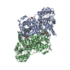 2w93C  2g1i S: Starting model for refinement C: citing same article ( |
|---|---|
| Similar structure data |

-Links
 PDBj
PDBj


- Assembly
| Deposited unit | 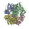
| ||||||||
|---|---|---|---|---|---|---|---|---|---|
| 1 | 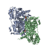
| ||||||||
| 2 | 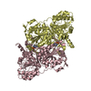
| ||||||||
| Unit cell |
|
-Components
| #1: Protein | Mass: 61716.762 Da / Num. of mol.: 4 / Source method: isolated from a natural source / Source: (natural) KLUYVEROMYCES LACTIS (yeast) / References: UniProt: Q12629, pyruvate decarboxylase#2: Chemical | ChemComp-TPP /   Mass: 425.314 Da / Num. of mol.: 4 / Source method: obtained synthetically / Formula: C12H19N4O7P2S Mass: 425.314 Da / Num. of mol.: 4 / Source method: obtained synthetically / Formula: C12H19N4O7P2S#3: Chemical | ChemComp-MG /   Mass: 24.305 Da / Num. of mol.: 4 / Source method: obtained synthetically / Formula: Mg Mass: 24.305 Da / Num. of mol.: 4 / Source method: obtained synthetically / Formula: Mg#4: Water | ChemComp-HOH / |  Mass: 18.015 Da / Num. of mol.: 1319 / Source method: isolated from a natural source / Formula: H2O Mass: 18.015 Da / Num. of mol.: 1319 / Source method: isolated from a natural source / Formula: H2O |
|---|
-Experimental details
-Experiment
| Experiment | Method: X-RAY DIFFRACTION / Number of used crystals: 1 |
|---|
- Sample preparation
| Crystal | Density Matthews: 2.59 Å3/Da / Density % sol: 52.59 % / Description: NONE |
|---|---|
| Crystal grow | Temperature: 281 K / Method: vapor diffusion, hanging drop / pH: 6.45 Details: 50MM MES, 1MM DTT, 5MM TDP, 5MM MAGNESIUM SULFATE, 10% PEG 2000, 10% PEG 8000, 2MG KLPDC/ML, PH 6.45, VAPOR DIFFUSION, HANGING DROP, TEMPERATURE 281K |
-Data collection
| Diffraction | Mean temperature: 100 K |
|---|---|
| Diffraction source | Source: SYNCHROTRON / Site: EMBL/DESY, HAMBURG  / Beamline: X11 / Wavelength: 0.8125 / Beamline: X11 / Wavelength: 0.8125 |
| Detector | Type: MARRESEARCH / Detector: CCD / Date: May 25, 2005 |
| Radiation | Protocol: SINGLE WAVELENGTH / Monochromatic (M) / Laue (L): M / Scattering type: x-ray |
| Radiation wavelength | Wavelength: 0.8125 Å / Relative weight: 1 |
| Reflection | Resolution: 1.95→48.45 Å / Num. obs: 155315 / % possible obs: 86.3 % / Observed criterion σ(I): 1 / Redundancy: 4.3 % / Rmerge(I) obs: 0.1 / Net I/σ(I): 11.5 |
| Reflection shell | Resolution: 1.95→2.06 Å / Rmerge(I) obs: 0.51 / Mean I/σ(I) obs: 2.5 / % possible all: 80.6 |
- Processing
| Software |
| ||||||||||||||||||||||||||||||||||||||||||||||||||||||||||||||||||||||||||||||||||||||||||||||||||||||||||||||||||||||||||||||||||||||||||||||||||||||||||||||||||||||||||||||||||||||
|---|---|---|---|---|---|---|---|---|---|---|---|---|---|---|---|---|---|---|---|---|---|---|---|---|---|---|---|---|---|---|---|---|---|---|---|---|---|---|---|---|---|---|---|---|---|---|---|---|---|---|---|---|---|---|---|---|---|---|---|---|---|---|---|---|---|---|---|---|---|---|---|---|---|---|---|---|---|---|---|---|---|---|---|---|---|---|---|---|---|---|---|---|---|---|---|---|---|---|---|---|---|---|---|---|---|---|---|---|---|---|---|---|---|---|---|---|---|---|---|---|---|---|---|---|---|---|---|---|---|---|---|---|---|---|---|---|---|---|---|---|---|---|---|---|---|---|---|---|---|---|---|---|---|---|---|---|---|---|---|---|---|---|---|---|---|---|---|---|---|---|---|---|---|---|---|---|---|---|---|---|---|---|---|
| Refinement | Method to determine structure: MOLECULAR REPLACEMENT Starting model: PDB ENTRY 2G1I 2g1i Resolution: 1.95→48.45 Å / Cor.coef. Fo:Fc: 0.951 / Cor.coef. Fo:Fc free: 0.92 / Cross valid method: THROUGHOUT / ESU R: 0.179 / ESU R Free: 0.163 / Stereochemistry target values: MAXIMUM LIKELIHOOD / Details: HYDROGENS HAVE BEEN ADDED IN THE RIDING POSITIONS.
| ||||||||||||||||||||||||||||||||||||||||||||||||||||||||||||||||||||||||||||||||||||||||||||||||||||||||||||||||||||||||||||||||||||||||||||||||||||||||||||||||||||||||||||||||||||||
| Solvent computation | Ion probe radii: 0.8 Å / Shrinkage radii: 0.8 Å / VDW probe radii: 1.2 Å / Solvent model: MASK | ||||||||||||||||||||||||||||||||||||||||||||||||||||||||||||||||||||||||||||||||||||||||||||||||||||||||||||||||||||||||||||||||||||||||||||||||||||||||||||||||||||||||||||||||||||||
| Displacement parameters | Biso mean: 22.08 Å2
| ||||||||||||||||||||||||||||||||||||||||||||||||||||||||||||||||||||||||||||||||||||||||||||||||||||||||||||||||||||||||||||||||||||||||||||||||||||||||||||||||||||||||||||||||||||||
| Refinement step | Cycle: LAST / Resolution: 1.95→48.45 Å
| ||||||||||||||||||||||||||||||||||||||||||||||||||||||||||||||||||||||||||||||||||||||||||||||||||||||||||||||||||||||||||||||||||||||||||||||||||||||||||||||||||||||||||||||||||||||
| Refine LS restraints |
|