 Movie
Movie Controller


+ Open data

- Basic information
| Entry | Database: PDB / ID: 6e8t | ||||||
|---|---|---|---|---|---|---|---|
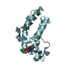
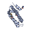
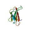
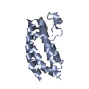
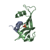
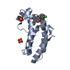
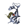
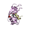
| Title | Structure of the Mango-III (A10U) aptamer bound to TO1-Biotin | ||||||
 Components Components | RNA (35-MER) | ||||||
 Keywords Keywords | RNA / Aptamer / fluorescence / G-quadruplex | ||||||
| Function / homology | Chem-HZG / : / RNA / RNA (> 10) Function and homology information Function and homology information | ||||||
| Biological species | synthetic construct (others) | ||||||
| Method |  X-RAY DIFFRACTION / SYNCHROTRON / MOLECULAR REPLACEMENT / Resolution: 2.9 Å X-RAY DIFFRACTION / SYNCHROTRON / MOLECULAR REPLACEMENT / Resolution: 2.9 Å | ||||||
 Authors Authors | Trachman, R.J. / Ferre-D'Amare, A.R. | ||||||
 Citation Citation | Journal: Nat. Chem. Biol. / Year: 2019 Title: Structure and functional reselection of the Mango-III fluorogenic RNA aptamer. Authors: Trachman 3rd., R.J. / Autour, A. / Jeng, S.C.Y. / Abdolahzadeh, A. / Andreoni, A. / Cojocaru, R. / Garipov, R. / Dolgosheina, E.V. / Knutson, J.R. / Ryckelynck, M. / Unrau, P.J. / Ferre-D'Amare, A.R. | ||||||
| History |
|
- Structure visualization
| Structure viewer | Molecule: MolmilJmol/JSmol |
|---|
- Downloads & links
-Download
| PDBx/mmCIF format | 6e8t.cif.gz | 113 KB | Display | PDBx/mmCIF format |
|---|---|---|---|---|
| PDB format | pdb6e8t.ent.gz | 69.5 KB | Display | PDB format |
| PDBx/mmJSON format | 6e8t.json.gz | Tree view | PDBx/mmJSON format | |
| Others |  Other downloads Other downloads |
-Validation report
| Arichive directory | https://data.pdbj.org/pub/pdb/validation_reports/e8/6e8tftp://data.pdbj.org/pub/pdb/validation_reports/e8/6e8t | HTTPS FTP |
|---|
-Related structure data
| Related structure data |  6e8sSC  6e8uC S: Starting model for refinement C: citing same article ( |
|---|---|
| Similar structure data |


-Links
 PDBj
PDBj


- Assembly
| Deposited unit | 
| ||||||||||||
|---|---|---|---|---|---|---|---|---|---|---|---|---|---|
| 1 | 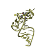
| ||||||||||||
| 2 | 
| ||||||||||||
| 3 | 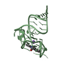
| ||||||||||||
| 4 | 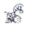
| ||||||||||||
| Unit cell |
| ||||||||||||
| Components on special symmetry positions |
|
-Components
| #1: RNA chain | Mass: 11649.898 Da / Num. of mol.: 4 / Source method: obtained synthetically / Details: synthetic / Source: (synth.) synthetic construct (others) #2: Chemical | ChemComp-HZG /   Mass: 348.461 Da / Num. of mol.: 4 / Source method: obtained synthetically / Formula: C21H20N2OS Mass: 348.461 Da / Num. of mol.: 4 / Source method: obtained synthetically / Formula: C21H20N2OS#3: Chemical | ChemComp-K /   Mass: 39.098 Da / Num. of mol.: 4 / Source method: obtained synthetically / Formula: K Mass: 39.098 Da / Num. of mol.: 4 / Source method: obtained synthetically / Formula: K#4: Chemical | ChemComp-NA / |   Mass: 22.990 Da / Num. of mol.: 1 / Source method: obtained synthetically / Formula: Na Mass: 22.990 Da / Num. of mol.: 1 / Source method: obtained synthetically / Formula: Na#5: Water | ChemComp-HOH / |  Mass: 18.015 Da / Num. of mol.: 4 / Source method: isolated from a natural source / Formula: H2O Mass: 18.015 Da / Num. of mol.: 4 / Source method: isolated from a natural source / Formula: H2O |
|---|
-Experimental details
-Experiment
| Experiment | Method: X-RAY DIFFRACTION / Number of used crystals: 1 |
|---|
- Sample preparation
| Crystal | Density Matthews: 2.84 Å3/Da / Density % sol: 56.73 % |
|---|---|
| Crystal grow | Temperature: 294 K / Method: vapor diffusion, sitting drop / Details: 1.6 M Ammonium Citrate, 3.0% Glycerol, 4% Acetone |
-Data collection
| Diffraction | Mean temperature: 100 K |
|---|---|
| Diffraction source | Source: SYNCHROTRON / Site: ALS  / Beamline: 5.0.2 / Wavelength: 1.105 Å / Beamline: 5.0.2 / Wavelength: 1.105 Å |
| Detector | Type: DECTRIS PILATUS3 6M / Detector: PIXEL / Date: Jun 14, 2017 |
| Radiation | Protocol: SINGLE WAVELENGTH / Monochromatic (M) / Laue (L): M / Scattering type: x-ray |
| Radiation wavelength | Wavelength: 1.105 Å / Relative weight: 1 |
| Reflection | Resolution: 2.9→45.97 Å / Num. obs: 18895 / % possible obs: 98.5 % / Redundancy: 2.3 % / Biso Wilson estimate: 77.73 Å2 / Rmerge(I) obs: 0.038 / Net I/σ(I): 10.3 |
| Reflection shell | Resolution: 2.9→3.03 Å / Redundancy: 2.2 % / Rmerge(I) obs: 0.91 / Num. unique obs: 1235 / % possible all: 99 |
- Processing
| Software |
| ||||||||||||||||||||||||||||||||||||||||||||||||||||||||||||||||||||||||||||||||||||||||||||||||||
|---|---|---|---|---|---|---|---|---|---|---|---|---|---|---|---|---|---|---|---|---|---|---|---|---|---|---|---|---|---|---|---|---|---|---|---|---|---|---|---|---|---|---|---|---|---|---|---|---|---|---|---|---|---|---|---|---|---|---|---|---|---|---|---|---|---|---|---|---|---|---|---|---|---|---|---|---|---|---|---|---|---|---|---|---|---|---|---|---|---|---|---|---|---|---|---|---|---|---|---|
| Refinement | Method to determine structure: MOLECULAR REPLACEMENT Starting model: 6E8S Resolution: 2.9→45.97 Å / SU ML: 0.5907 / Cross valid method: FREE R-VALUE / σ(F): 1.96 / Phase error: 32.2373
| ||||||||||||||||||||||||||||||||||||||||||||||||||||||||||||||||||||||||||||||||||||||||||||||||||
| Solvent computation | Shrinkage radii: 0.9 Å / VDW probe radii: 1.11 Å | ||||||||||||||||||||||||||||||||||||||||||||||||||||||||||||||||||||||||||||||||||||||||||||||||||
| Displacement parameters | Biso mean: 78.05 Å2 | ||||||||||||||||||||||||||||||||||||||||||||||||||||||||||||||||||||||||||||||||||||||||||||||||||
| Refinement step | Cycle: LAST / Resolution: 2.9→45.97 Å
| ||||||||||||||||||||||||||||||||||||||||||||||||||||||||||||||||||||||||||||||||||||||||||||||||||
| Refine LS restraints |
| ||||||||||||||||||||||||||||||||||||||||||||||||||||||||||||||||||||||||||||||||||||||||||||||||||
| LS refinement shell |
|
