 Movie
Movie Controller

[English] 日本語
 Yorodumi
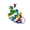
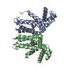
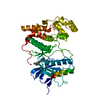
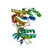
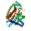
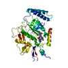
- PDB-6e66: Crystal structure of bacterial N-acetylglucosamine transferase NleB -
+ Open data

- Basic information
| Entry | Database: PDB / ID: 6.0E+66 | ||||||
|---|---|---|---|---|---|---|---|
| Title | Crystal structure of bacterial N-acetylglucosamine transferase NleB | ||||||
 Components Components | NleB | ||||||
 Keywords Keywords | TRANSFERASE / NleB / Bacterial effector / Arginine GlcNAcylation | ||||||
| Function / homology |  Function and homology information Function and homology informationprotein-arginine N-acetylglucosaminyltransferase activity / symbiont-mediated suppression of host programmed cell death / Transferases; Glycosyltransferases; Hexosyltransferases / manganese ion binding / toxin activity / symbiont-mediated suppression of host NF-kappaB cascade / host cell cytoplasm / extracellular region Similarity search - Function | ||||||
| Biological species |  | ||||||
| Method |  X-RAY DIFFRACTION / SYNCHROTRON / SAD / Resolution: 2.1 Å X-RAY DIFFRACTION / SYNCHROTRON / SAD / Resolution: 2.1 Å | ||||||
 Authors Authors | Yao, Q. / Zheng, Y.Q. / Shao, F. | ||||||
 Citation Citation | Journal: Mol.Cell / Year: 2019 Title: Structural and Functional Insights into Host Death Domains Inactivation by the Bacterial Arginine GlcNAcyltransferase Effector. Authors: Ding, J. / Pan, X. / Du, L. / Yao, Q. / Xue, J. / Yao, H. / Wang, D.C. / Li, S. / Shao, F. | ||||||
| History |
|
- Structure visualization
| Structure viewer | Molecule: MolmilJmol/JSmol |
|---|
- Downloads & links
-Download
| PDBx/mmCIF format | 6e66.cif.gz | 74.3 KB | Display | PDBx/mmCIF format |
|---|---|---|---|---|
| PDB format | pdb6e66.ent.gz | 53.4 KB | Display | PDB format |
| PDBx/mmJSON format | 6e66.json.gz | Tree view | PDBx/mmJSON format | |
| Others |  Other downloads Other downloads |
-Validation report
| Arichive directory | https://data.pdbj.org/pub/pdb/validation_reports/e6/6e66ftp://data.pdbj.org/pub/pdb/validation_reports/e6/6e66 | HTTPS FTP |
|---|
-Related structure data


-Links
 PDBj
PDBj- Assembly
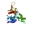
| Deposited unit | 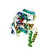
| ||||||||||
|---|---|---|---|---|---|---|---|---|---|---|---|
| 1 |
| ||||||||||
| Unit cell |
|
-Components
| #1: Protein | Mass: 38175.699 Da / Num. of mol.: 1 Source method: isolated from a genetically manipulated source Source: (gene. exp.) |
|---|---|
| #2: Chemical | ChemComp-EDO /   Mass: 62.068 Da / Num. of mol.: 1 / Source method: obtained synthetically / Formula: C2H6O2 Mass: 62.068 Da / Num. of mol.: 1 / Source method: obtained synthetically / Formula: C2H6O2 |
| #3: Water | ChemComp-HOH /  Mass: 18.015 Da / Num. of mol.: 84 / Source method: isolated from a natural source / Formula: H2O Mass: 18.015 Da / Num. of mol.: 84 / Source method: isolated from a natural source / Formula: H2O |
| Has protein modification | Y |
-Experimental details
-Experiment
| Experiment | Method: X-RAY DIFFRACTION / Number of used crystals: 1 |
|---|
- Sample preparation
| Crystal | Density Matthews: 2.23 Å3/Da / Density % sol: 44.82 % |
|---|---|
| Crystal grow | Temperature: 293 K / Method: vapor diffusion, hanging drop Details: 0.2 ammonium sulfate, 0.1 M sodium cacodylate trihydrate at pH 6.5 and 30% PEG 8000 |
-Data collection
| Diffraction | Mean temperature: 77.2 K |
|---|---|
| Diffraction source | Source: SYNCHROTRON / Site: SSRF  / Beamline: BL17B1 / Wavelength: 0.9791 Å / Beamline: BL17B1 / Wavelength: 0.9791 Å |
| Detector | Type: ADSC QUANTUM 315r / Detector: CCD / Date: Sep 12, 2011 |
| Radiation | Protocol: SINGLE WAVELENGTH / Monochromatic (M) / Laue (L): M / Scattering type: x-ray |
| Radiation wavelength | Wavelength: 0.9791 Å / Relative weight: 1 |
| Reflection | Resolution: 2.1→20 Å / Num. obs: 19964 / % possible obs: 97.94 % / Observed criterion σ(I): 3 / Redundancy: 6.5 % / Rmerge(I) obs: 0.214 / Net I/σ(I): 21.4 |
| Reflection shell | Resolution: 2.1→2.14 Å / Rmerge(I) obs: 0.439 / Num. unique obs: 1873 |
- Processing
| Software |
| ||||||||||||||||||||||||||||||||||||||||||||||||||||||||
|---|---|---|---|---|---|---|---|---|---|---|---|---|---|---|---|---|---|---|---|---|---|---|---|---|---|---|---|---|---|---|---|---|---|---|---|---|---|---|---|---|---|---|---|---|---|---|---|---|---|---|---|---|---|---|---|---|---|
| Refinement | Method to determine structure: SAD / Resolution: 2.1→19.744 Å / SU ML: 0.22 / Cross valid method: FREE R-VALUE / σ(F): 0.28 / Phase error: 24.51
| ||||||||||||||||||||||||||||||||||||||||||||||||||||||||
| Solvent computation | Shrinkage radii: 0.9 Å / VDW probe radii: 1.11 Å | ||||||||||||||||||||||||||||||||||||||||||||||||||||||||
| Refinement step | Cycle: LAST / Resolution: 2.1→19.744 Å
| ||||||||||||||||||||||||||||||||||||||||||||||||||||||||
| Refine LS restraints |
| ||||||||||||||||||||||||||||||||||||||||||||||||||||||||
| LS refinement shell |
|
