 Movie
Movie Controller


+ Open data

- Basic information
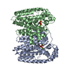
| Entry | Database: PDB / ID: 6shv | ||||||
|---|---|---|---|---|---|---|---|
| Title | T. cruzi FPPS in complex with 5-(4-fluorophenoxy)pyridin-2-amine | ||||||
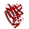
 Components Components | Farnesyl diphosphate synthase | ||||||
 Keywords Keywords | TRANSFERASE / farnesyl diphosphate synthase / sterol biosynthesis / farnesyl pyrophosphate / homodimer | ||||||
| Function / homology |  Function and homology information Function and homology informationtrans, trans-farnesyl diphosphate biosynthetic process / dimethylallyltranstransferase activity / (2E,6E)-farnesyl diphosphate synthase activity / metal ion binding / cytoplasm Similarity search - Function | ||||||
| Biological species |  | ||||||
| Method |  X-RAY DIFFRACTION / SYNCHROTRON / MOLECULAR REPLACEMENT / molecular replacement / Resolution: 1.808 Å X-RAY DIFFRACTION / SYNCHROTRON / MOLECULAR REPLACEMENT / molecular replacement / Resolution: 1.808 Å | ||||||
 Authors Authors | Petrick, J.K. / Muenzker, L. / Schleberger, C. / Cornaciu, I. / Clavel, D. / Marquez, J.A. / Jahnke, W. | ||||||
| Funding support | 1items
| ||||||
 Citation Citation | Journal: Thesis / Year: 2019 Title: Targeting farnesyl pyrophosphate synthase of Trypanosoma cruzi by fragment-based lead discovery Authors: Petrick, J.K. / Jahnke, W. | ||||||
| History |
|
- Structure visualization
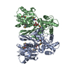
| Structure viewer | Molecule: MolmilJmol/JSmol |
|---|
- Downloads & links
-Download
| PDBx/mmCIF format | 6shv.cif.gz | 158.7 KB | Display | PDBx/mmCIF format |
|---|---|---|---|---|
| PDB format | pdb6shv.ent.gz | 122.6 KB | Display | PDB format |
| PDBx/mmJSON format | 6shv.json.gz | Tree view | PDBx/mmJSON format | |
| Others |  Other downloads Other downloads |
-Validation report
| Arichive directory | https://data.pdbj.org/pub/pdb/validation_reports/sh/6shvftp://data.pdbj.org/pub/pdb/validation_reports/sh/6shv | HTTPS FTP |
|---|
-Related structure data
| Related structure data |  6r04C  6r05C  6r06C  6r07C  6r08C  6r09C  6r0aC  6r0bC 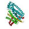 6r39C  6si5C 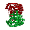 4dwgS S: Starting model for refinement C: citing same article ( |
|---|---|
| Similar structure data |


-Links
 PDBj
PDBj
- Assembly
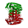
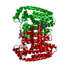
| Deposited unit | 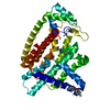
| ||||||||
|---|---|---|---|---|---|---|---|---|---|
| 1 | 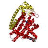
| ||||||||
| Unit cell |
|
-Components
| #1: Protein | Mass: 41359.574 Da / Num. of mol.: 1 Source method: isolated from a genetically manipulated source Source: (gene. exp.)  | ||||||||
|---|---|---|---|---|---|---|---|---|---|
| #2: Chemical |   Mass: 204.200 Da / Num. of mol.: 2 / Source method: obtained synthetically / Formula: C11H9FN2O / Feature type: SUBJECT OF INVESTIGATION Mass: 204.200 Da / Num. of mol.: 2 / Source method: obtained synthetically / Formula: C11H9FN2O / Feature type: SUBJECT OF INVESTIGATION#3: Chemical | ChemComp-ZN / |   Mass: 65.409 Da / Num. of mol.: 1 / Source method: obtained synthetically / Formula: Zn Mass: 65.409 Da / Num. of mol.: 1 / Source method: obtained synthetically / Formula: Zn#4: Chemical | ChemComp-SO4 / |   Mass: 96.063 Da / Num. of mol.: 1 / Source method: obtained synthetically / Formula: SO4 Mass: 96.063 Da / Num. of mol.: 1 / Source method: obtained synthetically / Formula: SO4#5: Water | ChemComp-HOH / |  Mass: 18.015 Da / Num. of mol.: 206 / Source method: isolated from a natural source / Formula: H2O Mass: 18.015 Da / Num. of mol.: 206 / Source method: isolated from a natural source / Formula: H2OHas ligand of interest | Y | |
-Experimental details
-Experiment
| Experiment | Method: X-RAY DIFFRACTION / Number of used crystals: 1 |
|---|
- Sample preparation
| Crystal | Density Matthews: 2.36 Å3/Da / Density % sol: 47.78 % / Mosaicity: 0.05 ° |
|---|---|
| Crystal grow | Temperature: 293.15 K / Method: vapor diffusion, sitting drop / pH: 6.5 Details: 80 mM MES: 4.0 mM ZnSO4: 12.36 % PEG MME 550: 11.57% glycerol |
-Data collection
| Diffraction | Mean temperature: 100 K / Serial crystal experiment: N |
|---|---|
| Diffraction source | Source: SYNCHROTRON / Site: ESRF  / Beamline: ID30B / Wavelength: 0.97625 Å / Beamline: ID30B / Wavelength: 0.97625 Å |
| Detector | Type: DECTRIS PILATUS 6M / Detector: PIXEL / Date: Apr 20, 2018 |
| Radiation | Protocol: SINGLE WAVELENGTH / Monochromatic (M) / Laue (L): M / Scattering type: x-ray |
| Radiation wavelength | Wavelength: 0.97625 Å / Relative weight: 1 |
| Reflection | Resolution: 1.808→44.978 Å / Num. obs: 38338 / % possible obs: 100 % / Redundancy: 12.3 % / Biso Wilson estimate: 27.48 Å2 / CC1/2: 0.998 / Rmerge(I) obs: 0.099 / Rpim(I) all: 0.03 / Rrim(I) all: 0.103 / Rsym value: 0.099 / Net I/σ(I): 15.1 |
| Reflection shell | Resolution: 1.808→1.839 Å / Redundancy: 12.2 % / Rmerge(I) obs: 1.223 / Mean I/σ(I) obs: 2.1 / Num. unique obs: 1850 / CC1/2: 0.705 / Rpim(I) all: 0.361 / Rrim(I) all: 1.277 / Rsym value: 1.223 / % possible all: 100 |
-Phasing
| Phasing | Method: molecular replacement | ||||||
|---|---|---|---|---|---|---|---|
| Phasing MR | Model details: Phaser MODE: MR_TRA
|
- Processing
| Software |
| ||||||||||||||||||||||||||||||||||||||||||||||||||||||||||||||||||||||||||||||||||||||||||||||||||||||||||||
|---|---|---|---|---|---|---|---|---|---|---|---|---|---|---|---|---|---|---|---|---|---|---|---|---|---|---|---|---|---|---|---|---|---|---|---|---|---|---|---|---|---|---|---|---|---|---|---|---|---|---|---|---|---|---|---|---|---|---|---|---|---|---|---|---|---|---|---|---|---|---|---|---|---|---|---|---|---|---|---|---|---|---|---|---|---|---|---|---|---|---|---|---|---|---|---|---|---|---|---|---|---|---|---|---|---|---|---|---|---|
| Refinement | Method to determine structure: MOLECULAR REPLACEMENT Starting model: 4DWG Resolution: 1.808→44.978 Å / Cor.coef. Fo:Fc: 0.957 / Cor.coef. Fo:Fc free: 0.947 / SU R Cruickshank DPI: 0.103 / Cross valid method: THROUGHOUT / σ(F): 0 / SU R Blow DPI: 0.109 / SU Rfree Blow DPI: 0.107 / SU Rfree Cruickshank DPI: 0.103
| ||||||||||||||||||||||||||||||||||||||||||||||||||||||||||||||||||||||||||||||||||||||||||||||||||||||||||||
| Displacement parameters | Biso max: 103.34 Å2 / Biso mean: 34.05 Å2 / Biso min: 15.75 Å2
| ||||||||||||||||||||||||||||||||||||||||||||||||||||||||||||||||||||||||||||||||||||||||||||||||||||||||||||
| Refine analyze | Luzzati coordinate error obs: 0.23 Å | ||||||||||||||||||||||||||||||||||||||||||||||||||||||||||||||||||||||||||||||||||||||||||||||||||||||||||||
| Refinement step | Cycle: final / Resolution: 1.808→44.978 Å
| ||||||||||||||||||||||||||||||||||||||||||||||||||||||||||||||||||||||||||||||||||||||||||||||||||||||||||||
| Refine LS restraints |
| ||||||||||||||||||||||||||||||||||||||||||||||||||||||||||||||||||||||||||||||||||||||||||||||||||||||||||||
| LS refinement shell | Resolution: 1.808→1.839 Å / Rfactor Rfree error: 0
| ||||||||||||||||||||||||||||||||||||||||||||||||||||||||||||||||||||||||||||||||||||||||||||||||||||||||||||
| Refinement TLS params. | Method: refined / Origin x: -9.2047 Å / Origin y: -17.8696 Å / Origin z: -20.6741 Å
| ||||||||||||||||||||||||||||||||||||||||||||||||||||||||||||||||||||||||||||||||||||||||||||||||||||||||||||
| Refinement TLS group | Selection details: { A|* } |
