 Movie
Movie Controller

[English] 日本語
 Yorodumi
- PDB-3mav: Crystal structure of Plasmodium vivax putative farnesyl pyrophosp... -
+ Open data

- Basic information
| Entry | Database: PDB / ID: 3mav | |||||||||
|---|---|---|---|---|---|---|---|---|---|---|
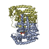
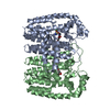
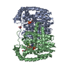
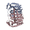
| Title | Crystal structure of Plasmodium vivax putative farnesyl pyrophosphate synthase (Pv092040) | |||||||||
 Components Components | Farnesyl pyrophosphate synthase | |||||||||
 Keywords Keywords | TRANSFERASE / PLASMODIUM VIVAX FARNESYL PYROPHOSPHATE SYNTHASE / PV092040 / STRUCTURAL GENOMICS / STRUCTURAL GENOMICS CONSORTIUM / SGC / Isoprene biosynthesis | |||||||||
| Function / homology |  Function and homology information Function and homology informationtrans, trans-farnesyl diphosphate biosynthetic process / dimethylallyltranstransferase activity / (2E,6E)-farnesyl diphosphate synthase activity / metal ion binding / cytoplasm Similarity search - Function | |||||||||
| Biological species |  | |||||||||
| Method |  X-RAY DIFFRACTION / MOLECULAR REPLACEMENT / Resolution: 2.1 Å X-RAY DIFFRACTION / MOLECULAR REPLACEMENT / Resolution: 2.1 Å | |||||||||
 Authors Authors | Dong, A. / Dunford, J. / Lew, J. / Wernimont, A.K. / Ren, H. / Zhao, Y. / Koeieradzki, I. / Opperman, U. / Sundstrom, M. / Weigelt, J. ...Dong, A. / Dunford, J. / Lew, J. / Wernimont, A.K. / Ren, H. / Zhao, Y. / Koeieradzki, I. / Opperman, U. / Sundstrom, M. / Weigelt, J. / Edwards, A.M. / Arrowsmith, C.H. / Bochkarev, A. / Hui, R. / Artz, J.D. / Structural Genomics Consortium (SGC) | |||||||||
 Citation Citation | Journal: J.Biol.Chem. / Year: 2011 Title: Molecular characterization of a novel geranylgeranyl pyrophosphate synthase from Plasmodium parasites. Authors: Artz, J.D. / Wernimont, A.K. / Dunford, J.E. / Schapira, M. / Dong, A. / Zhao, Y. / Lew, J. / Russell, R.G. / Ebetino, F.H. / Oppermann, U. / Hui, R. | |||||||||
| History |
|
- Structure visualization
| Structure viewer | Molecule: MolmilJmol/JSmol |
|---|
- Downloads & links
-Download
| PDBx/mmCIF format | 3mav.cif.gz | 289.5 KB | Display | PDBx/mmCIF format |
|---|---|---|---|---|
| PDB format | pdb3mav.ent.gz | 230.8 KB | Display | PDB format |
| PDBx/mmJSON format | 3mav.json.gz | Tree view | PDBx/mmJSON format | |
| Others |  Other downloads Other downloads |
-Validation report
| Arichive directory | https://data.pdbj.org/pub/pdb/validation_reports/ma/3mavftp://data.pdbj.org/pub/pdb/validation_reports/ma/3mav | HTTPS FTP |
|---|
-Related structure data
| Related structure data |  3ldwC  3ph7C  1yhkS S: Starting model for refinement C: citing same article ( |
|---|---|
| Similar structure data |


-Links
 PDBj
PDBj
- Assembly
| Deposited unit | 
| ||||||||
|---|---|---|---|---|---|---|---|---|---|
| 1 | 
| ||||||||
| 2 | 
| ||||||||
| Unit cell |
|
-Components
| #1: Protein | Mass: 46084.555 Da / Num. of mol.: 4 Source method: isolated from a genetically manipulated source Source: (gene. exp.) Strain: SALVADOR I / Gene: PVX_092040 / Plasmid: P15-TEV-LIC DERIVED FROM PET15 / Production host:  #2: Chemical | ChemComp-SO4 /   Mass: 96.063 Da / Num. of mol.: 9 / Source method: obtained synthetically / Formula: SO4 Mass: 96.063 Da / Num. of mol.: 9 / Source method: obtained synthetically / Formula: SO4#3: Water | ChemComp-HOH / |  Mass: 18.015 Da / Num. of mol.: 370 / Source method: isolated from a natural source / Formula: H2O Mass: 18.015 Da / Num. of mol.: 370 / Source method: isolated from a natural source / Formula: H2O |
|---|
-Experimental details
-Experiment
| Experiment | Method: X-RAY DIFFRACTION / Number of used crystals: 1 |
|---|
- Sample preparation
| Crystal | Density Matthews: 2.2 Å3/Da / Density % sol: 44.13 % |
|---|---|
| Crystal grow | Temperature: 297 K / Method: vapor diffusion, hanging drop / pH: 8.5 Details: 22% PEG 3350, 200 MM LI2SO4, 100 MM TRIS pH 8.5, VAPOR DIFFUSION, HANGING DROP, temperature 297K |
-Data collection
| Diffraction | Mean temperature: 100 K |
|---|---|
| Diffraction source | Source: ROTATING ANODE / Type: RIGAKU FR-E SUPERBRIGHT / Wavelength: 1.54 Å |
| Detector | Type: RIGAKU RAXIS IV++ / Detector: IMAGE PLATE / Date: Sep 12, 2006 / Details: M |
| Radiation | Protocol: SINGLE WAVELENGTH / Monochromatic (M) / Laue (L): M / Scattering type: x-ray |
| Radiation wavelength | Wavelength: 1.54 Å / Relative weight: 1 |
| Reflection | Resolution: 2.1→50 Å / Num. all: 92263 / Num. obs: 92263 / % possible obs: 99.1 % / Observed criterion σ(F): 0 / Observed criterion σ(I): 0 / Redundancy: 4.7 % / Biso Wilson estimate: 43.16 Å2 / Rmerge(I) obs: 0.054 / Rsym value: 0.054 / Net I/σ(I): 17.5 |
| Reflection shell | Resolution: 2.1→2.14 Å / Redundancy: 4.4 % / Rmerge(I) obs: 0.564 / Mean I/σ(I) obs: 2.53 / Num. unique all: 4429 / Rsym value: 0.564 / % possible all: 96.6 |
- Processing
| Software |
| ||||||||||||||||||||||||||||||||||||||||||||||||||||||||||||
|---|---|---|---|---|---|---|---|---|---|---|---|---|---|---|---|---|---|---|---|---|---|---|---|---|---|---|---|---|---|---|---|---|---|---|---|---|---|---|---|---|---|---|---|---|---|---|---|---|---|---|---|---|---|---|---|---|---|---|---|---|---|
| Refinement | Method to determine structure: MOLECULAR REPLACEMENT Starting model: PDB ENTRY 1YHK Resolution: 2.1→20.99 Å / Cross valid method: THROUGHOUT / σ(F): 0 / σ(I): 0 / Stereochemistry target values: Engh & Huber
| ||||||||||||||||||||||||||||||||||||||||||||||||||||||||||||
| Displacement parameters | Biso mean: 46.29 Å2
| ||||||||||||||||||||||||||||||||||||||||||||||||||||||||||||
| Refine analyze | Luzzati coordinate error obs: 0.337 Å | ||||||||||||||||||||||||||||||||||||||||||||||||||||||||||||
| Refinement step | Cycle: LAST / Resolution: 2.1→20.99 Å
| ||||||||||||||||||||||||||||||||||||||||||||||||||||||||||||
| Refine LS restraints |
| ||||||||||||||||||||||||||||||||||||||||||||||||||||||||||||
| LS refinement shell | Resolution: 2.1→2.14 Å / Total num. of bins used: 20
|
