 Movie
Movie Controller

[English] 日本語
 Yorodumi
- PDB-5hn9: Crystal structure of Plasmodium vivax geranylgeranylpyrophosphate... -
+ Open data

- Basic information
| Entry | Database: PDB / ID: 5hn9 | ||||||
|---|---|---|---|---|---|---|---|
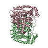
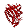
| Title | Crystal structure of Plasmodium vivax geranylgeranylpyrophosphate synthase complexed with BPH-1186 | ||||||
 Components Components | Farnesyl pyrophosphate synthase, putative | ||||||
 Keywords Keywords | TRANSFERASE/TRANSFERASE INHIBITOR / ALL HELICES / TRANSFERASE / TRANSFERASE-TRANSFERASE INHIBITOR COMPLEX | ||||||
| Function / homology |  Function and homology information Function and homology informationtrans, trans-farnesyl diphosphate biosynthetic process / dimethylallyltranstransferase activity / (2E,6E)-farnesyl diphosphate synthase activity / metal ion binding / cytoplasm Similarity search - Function | ||||||
| Biological species |  | ||||||
| Method |  X-RAY DIFFRACTION / SYNCHROTRON / MOLECULAR REPLACEMENT / Resolution: 2.12 Å X-RAY DIFFRACTION / SYNCHROTRON / MOLECULAR REPLACEMENT / Resolution: 2.12 Å | ||||||
 Authors Authors | Liu, Y.-L. / Zhang, Y. / OIdfield, E. | ||||||
| Funding support |  United States, 1items United States, 1items
| ||||||
 Citation Citation | Journal: Biochemistry / Year: 2016 Title: Dynamic Structure and Inhibition of a Malaria Drug Target: Geranylgeranyl Diphosphate Synthase. Authors: G Ricci, C. / Liu, Y.L. / Zhang, Y. / Wang, Y. / Zhu, W. / Oldfield, E. / McCammon, J.A. | ||||||
| History |
|
- Structure visualization
| Structure viewer | Molecule: MolmilJmol/JSmol |
|---|
- Downloads & links
-Download
| PDBx/mmCIF format | 5hn9.cif.gz | 571.9 KB | Display | PDBx/mmCIF format |
|---|---|---|---|---|
| PDB format | pdb5hn9.ent.gz | 471.9 KB | Display | PDB format |
| PDBx/mmJSON format | 5hn9.json.gz | Tree view | PDBx/mmJSON format | |
| Others |  Other downloads Other downloads |
-Validation report
| Arichive directory | https://data.pdbj.org/pub/pdb/validation_reports/hn/5hn9ftp://data.pdbj.org/pub/pdb/validation_reports/hn/5hn9 | HTTPS FTP |
|---|
-Related structure data
| Related structure data |  5hn7C  5hn8C  5hnaC  3mys C: citing same article ( S: Starting model for refinement |
|---|---|
| Similar structure data |


-Links
 PDBj
PDBj
- Assembly
| Deposited unit | 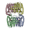
| ||||||||
|---|---|---|---|---|---|---|---|---|---|
| 1 | 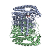
| ||||||||
| 2 | 
| ||||||||
| Unit cell |
|
-Components
| #1: Protein | Mass: 43784.188 Da / Num. of mol.: 4 Source method: isolated from a genetically manipulated source Source: (gene. exp.) Strain: Salvador I / Gene: PVX_092040 / Plasmid: P15-TEV-LIC / Production host:  #2: Chemical | ChemComp-04W /   Mass: 429.506 Da / Num. of mol.: 4 / Source method: obtained synthetically / Formula: C24H31NO6 Mass: 429.506 Da / Num. of mol.: 4 / Source method: obtained synthetically / Formula: C24H31NO6#3: Water | ChemComp-HOH / |  Mass: 18.015 Da / Num. of mol.: 406 / Source method: isolated from a natural source / Formula: H2O Mass: 18.015 Da / Num. of mol.: 406 / Source method: isolated from a natural source / Formula: H2OSequence details | THE AUTHORS STATE THAT THEY HAVE DETERMINED | |
|---|
-Experimental details
-Experiment
| Experiment | Method: X-RAY DIFFRACTION / Number of used crystals: 1 |
|---|
- Sample preparation
| Crystal | Density Matthews: 2.31 Å3/Da / Density % sol: 46.79 % |
|---|---|
| Crystal grow | Temperature: 291 K / Method: vapor diffusion, hanging drop / pH: 8.5 Details: 100MM TRIS, 200MM LITHIUM SULFATE, 22% PEG 3,350, PH 8.5 |
-Data collection
| Diffraction | Mean temperature: 293 K |
|---|---|
| Diffraction source | Source: SYNCHROTRON / Site: APS / Beamline: 21-ID-F / Wavelength: 0.97872 Å |
| Detector | Type: MARMOSAIC 225 mm CCD / Detector: CCD / Date: Dec 21, 2010 |
| Radiation | Protocol: SINGLE WAVELENGTH / Monochromatic (M) / Laue (L): M / Scattering type: x-ray |
| Radiation wavelength | Wavelength: 0.97872 Å / Relative weight: 1 |
| Reflection | Resolution: 2.1→50 Å / Num. obs: 91912 / % possible obs: 99.8 % / Observed criterion σ(I): 1 / Redundancy: 7.5 % / Rmerge(I) obs: 0.094 / Net I/σ(I): 9.9 |
| Reflection shell | Resolution: 2.1→2.14 Å / Redundancy: 5.4 % / Rmerge(I) obs: 0.581 / % possible all: 99.5 |
- Processing
| Software |
| ||||||||||||||||||||||||||||||||||||||||||||||||||||||||||||||||||||||||||||||||||||||||||||||||||||||||||||||||||||||||||||||||||||||||||||||||||||||||||||||||||||||||||||||||||||||
|---|---|---|---|---|---|---|---|---|---|---|---|---|---|---|---|---|---|---|---|---|---|---|---|---|---|---|---|---|---|---|---|---|---|---|---|---|---|---|---|---|---|---|---|---|---|---|---|---|---|---|---|---|---|---|---|---|---|---|---|---|---|---|---|---|---|---|---|---|---|---|---|---|---|---|---|---|---|---|---|---|---|---|---|---|---|---|---|---|---|---|---|---|---|---|---|---|---|---|---|---|---|---|---|---|---|---|---|---|---|---|---|---|---|---|---|---|---|---|---|---|---|---|---|---|---|---|---|---|---|---|---|---|---|---|---|---|---|---|---|---|---|---|---|---|---|---|---|---|---|---|---|---|---|---|---|---|---|---|---|---|---|---|---|---|---|---|---|---|---|---|---|---|---|---|---|---|---|---|---|---|---|---|---|
| Refinement | Method to determine structure: MOLECULAR REPLACEMENT Starting model: PDB ENTRY 3MYS 3mys Resolution: 2.12→47.94 Å / Cor.coef. Fo:Fc: 0.95 / Cor.coef. Fo:Fc free: 0.907 / SU B: 11.192 / SU ML: 0.136 / Cross valid method: THROUGHOUT / σ(F): 0 / ESU R Free: 0.198 / Stereochemistry target values: MAXIMUM LIKELIHOOD / Details: HYDROGENS HAVE BEEN ADDED IN THE RIDING
| ||||||||||||||||||||||||||||||||||||||||||||||||||||||||||||||||||||||||||||||||||||||||||||||||||||||||||||||||||||||||||||||||||||||||||||||||||||||||||||||||||||||||||||||||||||||
| Solvent computation | Ion probe radii: 0.8 Å / Shrinkage radii: 0.8 Å / VDW probe radii: 1.4 Å / Solvent model: MASK | ||||||||||||||||||||||||||||||||||||||||||||||||||||||||||||||||||||||||||||||||||||||||||||||||||||||||||||||||||||||||||||||||||||||||||||||||||||||||||||||||||||||||||||||||||||||
| Displacement parameters | Biso mean: 33.03 Å2
| ||||||||||||||||||||||||||||||||||||||||||||||||||||||||||||||||||||||||||||||||||||||||||||||||||||||||||||||||||||||||||||||||||||||||||||||||||||||||||||||||||||||||||||||||||||||
| Refinement step | Cycle: LAST / Resolution: 2.12→47.94 Å
| ||||||||||||||||||||||||||||||||||||||||||||||||||||||||||||||||||||||||||||||||||||||||||||||||||||||||||||||||||||||||||||||||||||||||||||||||||||||||||||||||||||||||||||||||||||||
| Refine LS restraints |
|