 Movie
Movie Controller

[English] 日本語
 Yorodumi
- PDB-6ac0: Crystal structure of TRADD death domain GlcNAcylated by EPEC effe... -
+ Open data

- Basic information
| Entry | Database: PDB / ID: 6ac0 | ||||||
|---|---|---|---|---|---|---|---|
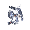
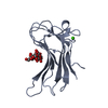
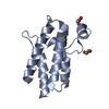
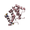
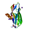
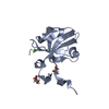
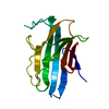
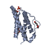
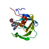
| Title | Crystal structure of TRADD death domain GlcNAcylated by EPEC effector NleB | ||||||
 Components Components | Tumor necrosis factor receptor type 1-associated DEATH domain protein | ||||||
 Keywords Keywords | SIGNALING PROTEIN / apoptosis | ||||||
| Function / homology |  Function and homology information Function and homology informationtumor necrosis factor receptor superfamily complex / death domain binding / Defective RIPK1-mediated regulated necrosis / TRAIL-activated apoptotic signaling pathway / positive regulation of hair follicle development / Regulation by c-FLIP / CASP8 activity is inhibited / Dimerization of procaspase-8 / TNF signaling / Caspase activation via Death Receptors in the presence of ligand ...tumor necrosis factor receptor superfamily complex / death domain binding / Defective RIPK1-mediated regulated necrosis / TRAIL-activated apoptotic signaling pathway / positive regulation of hair follicle development / Regulation by c-FLIP / CASP8 activity is inhibited / Dimerization of procaspase-8 / TNF signaling / Caspase activation via Death Receptors in the presence of ligand / death-inducing signaling complex / transmembrane receptor protein tyrosine kinase adaptor activity / TNFR1-induced proapoptotic signaling / RIPK1-mediated regulated necrosis / extrinsic apoptotic signaling pathway via death domain receptors / extrinsic apoptotic signaling pathway / canonical NF-kappaB signal transduction / signaling adaptor activity / positive regulation of interferon-beta production / tumor necrosis factor-mediated signaling pathway / negative regulation of canonical NF-kappaB signal transduction / TNFR1-induced NF-kappa-B signaling pathway / Regulation of TNFR1 signaling / Regulation of necroptotic cell death / cellular response to tumor necrosis factor / kinase binding / cytoplasmic side of plasma membrane / protein polyubiquitination / positive regulation of inflammatory response / cytoskeleton / protein-macromolecule adaptor activity / positive regulation of canonical NF-kappaB signal transduction / signaling receptor complex / positive regulation of cell migration / positive regulation of apoptotic process / apoptotic process / signal transduction / identical protein binding / nucleus / plasma membrane / cytoplasm / cytosol Similarity search - Function | ||||||
| Biological species |  Homo sapiens (human) Homo sapiens (human) | ||||||
| Method |  X-RAY DIFFRACTION / SYNCHROTRON / MOLECULAR REPLACEMENT / Resolution: 1.449 Å X-RAY DIFFRACTION / SYNCHROTRON / MOLECULAR REPLACEMENT / Resolution: 1.449 Å | ||||||
 Authors Authors | Ding, J. / Shao, F. | ||||||
 Citation Citation | Journal: Mol.Cell / Year: 2019 Title: Structural and Functional Insights into Host Death Domains Inactivation by the Bacterial Arginine GlcNAcyltransferase Effector. Authors: Ding, J. / Pan, X. / Du, L. / Yao, Q. / Xue, J. / Yao, H. / Wang, D.C. / Li, S. / Shao, F. | ||||||
| History |
|
- Structure visualization
| Structure viewer | Molecule: MolmilJmol/JSmol |
|---|
- Downloads & links
-Download
| PDBx/mmCIF format | 6ac0.cif.gz | 63.1 KB | Display | PDBx/mmCIF format |
|---|---|---|---|---|
| PDB format | pdb6ac0.ent.gz | 44.7 KB | Display | PDB format |
| PDBx/mmJSON format | 6ac0.json.gz | Tree view | PDBx/mmJSON format | |
| Others |  Other downloads Other downloads |
-Validation report
| Arichive directory | https://data.pdbj.org/pub/pdb/validation_reports/ac/6ac0ftp://data.pdbj.org/pub/pdb/validation_reports/ac/6ac0 | HTTPS FTP |
|---|
-Related structure data
| Related structure data |  6ac5C  6aciC 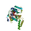 6e66C  3oq9S S: Starting model for refinement C: citing same article ( |
|---|---|
| Similar structure data |

-Links
 PDBj
PDBj


- Assembly
| Deposited unit | 
| ||||||||
|---|---|---|---|---|---|---|---|---|---|
| 1 |
| ||||||||
| Unit cell |
|
-Components
| #1: Protein | Mass: 13325.075 Da / Num. of mol.: 1 Source method: isolated from a genetically manipulated source Source: (gene. exp.) Homo sapiens (human) / Gene: TRADD / Plasmid: pGEX6p / Production host:  |
|---|---|
| #2: Sugar | ChemComp-NAG /   Type: D-saccharide, beta linking / Mass: 221.208 Da / Num. of mol.: 1 Type: D-saccharide, beta linking / Mass: 221.208 Da / Num. of mol.: 1Source method: isolated from a genetically manipulated source Formula: C8H15NO6 / Feature type: SUBJECT OF INVESTIGATION |
| #3: Water | ChemComp-HOH /  Mass: 18.015 Da / Num. of mol.: 104 / Source method: isolated from a natural source / Formula: H2O Mass: 18.015 Da / Num. of mol.: 104 / Source method: isolated from a natural source / Formula: H2O |
| Has protein modification | Y |
-Experimental details
-Experiment
| Experiment | Method: X-RAY DIFFRACTION / Number of used crystals: 1 |
|---|
- Sample preparation
| Crystal | Density Matthews: 2.03 Å3/Da / Density % sol: 39.45 % |
|---|---|
| Crystal grow | Temperature: 293 K / Method: vapor diffusion, sitting drop / pH: 6 / Details: 1.6 M Na/KPO4, pH 6.0 |
-Data collection
| Diffraction | Mean temperature: 93 K |
|---|---|
| Diffraction source | Source: SYNCHROTRON / Site: SSRF  / Beamline: BL17U / Wavelength: 0.97899 Å / Beamline: BL17U / Wavelength: 0.97899 Å |
| Detector | Type: ADSC QUANTUM 315r / Detector: CCD / Date: Nov 9, 2013 |
| Radiation | Protocol: SINGLE WAVELENGTH / Monochromatic (M) / Laue (L): M / Scattering type: x-ray |
| Radiation wavelength | Wavelength: 0.97899 Å / Relative weight: 1 |
| Reflection | Resolution: 1.449→28.6 Å / Num. obs: 20061 / % possible obs: 96.7 % / Redundancy: 4.6 % / Rmerge(I) obs: 0.111 / Net I/σ(I): 8.1 |
| Reflection shell | Resolution: 1.449→1.53 Å / Redundancy: 5.3 % / Rmerge(I) obs: 0.349 / Mean I/σ(I) obs: 3.8 / Num. unique obs: 2913 / % possible all: 99.3 |
- Processing
| Software |
| ||||||||||||||||||||||||||||||||||||||||||||||||||||||||
|---|---|---|---|---|---|---|---|---|---|---|---|---|---|---|---|---|---|---|---|---|---|---|---|---|---|---|---|---|---|---|---|---|---|---|---|---|---|---|---|---|---|---|---|---|---|---|---|---|---|---|---|---|---|---|---|---|---|
| Refinement | Method to determine structure: MOLECULAR REPLACEMENT Starting model: 3OQ9 Resolution: 1.449→28.435 Å / SU ML: 0.17 / Cross valid method: FREE R-VALUE / σ(F): 1.36 / Phase error: 20.74
| ||||||||||||||||||||||||||||||||||||||||||||||||||||||||
| Solvent computation | Shrinkage radii: 0.9 Å / VDW probe radii: 1.11 Å | ||||||||||||||||||||||||||||||||||||||||||||||||||||||||
| Refinement step | Cycle: LAST / Resolution: 1.449→28.435 Å
| ||||||||||||||||||||||||||||||||||||||||||||||||||||||||
| Refine LS restraints |
| ||||||||||||||||||||||||||||||||||||||||||||||||||||||||
| LS refinement shell |
|
