 Movie
Movie Controller

[English] 日本語
 Yorodumi
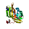
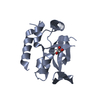
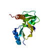
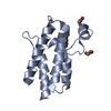
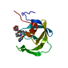
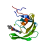
- PDB-1lmi: 1.5 ANGSTROM RESOLUTION CRYSTAL STRUCTURE OF A SECRETED PROTEIN F... -
+ Open data

- Basic information
| Entry | Database: PDB / ID: 1lmi | ||||||
|---|---|---|---|---|---|---|---|
| Title | 1.5 ANGSTROM RESOLUTION CRYSTAL STRUCTURE OF A SECRETED PROTEIN FROM MYCOBACTERIUM TUBERCULOSIS-MPT63 | ||||||
 Components Components | Immunogenic protein MPT63/MPB63 | ||||||
 Keywords Keywords | IMMUNE SYSTEM / beta-sandwich / Structural Genomics / PSI / Protein Structure Initiative / TB Structural Genomics Consortium / TBSGC | ||||||
| Function / homology |  Function and homology information Function and homology informationnucleotidyltransferase activity / peptidoglycan-based cell wall / : / extracellular region Similarity search - Function | ||||||
| Biological species |   Mycobacterium tuberculosis (bacteria) Mycobacterium tuberculosis (bacteria) | ||||||
| Method |  X-RAY DIFFRACTION / SYNCHROTRON / AB INITIO / Resolution: 1.5 Å X-RAY DIFFRACTION / SYNCHROTRON / AB INITIO / Resolution: 1.5 Å | ||||||
 Authors Authors | Goulding, C.W. / Parseghian, A. / Sawaya, M.R. / Cascio, D. / Apostol, M. / Gennaro, M.L. / Eisenberg, D. / TB Structural Genomics Consortium (TBSGC) | ||||||
 Citation Citation | Journal: Protein Sci. / Year: 2002 Title: Crystal structure of a major secreted protein of Mycobacterium tuberculosis-MPT63 at 1.5-A resolution Authors: Goulding, C.W. / Parseghian, A. / Sawaya, M.R. / Cascio, D. / Apostol, M. / Gennaro, M.L. / Eisenberg, D. | ||||||
| History |
|
- Structure visualization
| Structure viewer | Molecule: MolmilJmol/JSmol |
|---|
- Downloads & links
-Download
| PDBx/mmCIF format | 1lmi.cif.gz | 40.1 KB | Display | PDBx/mmCIF format |
|---|---|---|---|---|
| PDB format | pdb1lmi.ent.gz | 28 KB | Display | PDB format |
| PDBx/mmJSON format | 1lmi.json.gz | Tree view | PDBx/mmJSON format | |
| Others |  Other downloads Other downloads |
-Validation report
| Arichive directory | https://data.pdbj.org/pub/pdb/validation_reports/lm/1lmiftp://data.pdbj.org/pub/pdb/validation_reports/lm/1lmi | HTTPS FTP |
|---|
-Related structure data
| Similar structure data | |
|---|---|
| Other databases |


-Links
 PDBj
PDBj- Assembly
| Deposited unit | 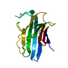
| |||||||||
|---|---|---|---|---|---|---|---|---|---|---|
| 1 |
| |||||||||
| Unit cell |
| |||||||||
| Components on special symmetry positions |
|
-Components
| #1: Protein | Mass: 13749.392 Da / Num. of mol.: 1 Source method: isolated from a genetically manipulated source Source: (gene. exp.) Mycobacterium tuberculosis (bacteria) / Tissue: H37Rv / Gene: Rv1926c, MPT63 / Plasmid: pQE30 / Species (production host): Escherichia coli / Production host: |
|---|---|
| #2: Water | ChemComp-HOH /  Mass: 18.015 Da / Num. of mol.: 172 / Source method: isolated from a natural source / Formula: H2O Mass: 18.015 Da / Num. of mol.: 172 / Source method: isolated from a natural source / Formula: H2O |
-Experimental details
-Experiment
| Experiment | Method: X-RAY DIFFRACTION / Number of used crystals: 1 |
|---|
- Sample preparation
| Crystal | Density Matthews: 1.65 Å3/Da / Density % sol: 25.6 % | ||||||||||||||||||||||||||||||
|---|---|---|---|---|---|---|---|---|---|---|---|---|---|---|---|---|---|---|---|---|---|---|---|---|---|---|---|---|---|---|---|
| Crystal grow | Temperature: 298 K / Method: vapor diffusion, hanging drop / pH: 7 Details: 26.5% PEG4000, 0.1M Tris/HCl, 15% glycerol, pH 7.0, VAPOR DIFFUSION, HANGING DROP, temperature 298K | ||||||||||||||||||||||||||||||
| Crystal grow | *PLUS | ||||||||||||||||||||||||||||||
| Components of the solutions | *PLUS
|
-Data collection
| Diffraction | Mean temperature: 190 K |
|---|---|
| Diffraction source | Source: SYNCHROTRON / Site: NSLS  / Beamline: X8C / Wavelength: 0.9792 Å / Beamline: X8C / Wavelength: 0.9792 Å |
| Detector | Type: ADSC QUANTUM 4 / Detector: CCD / Date: Oct 10, 2000 |
| Radiation | Monochromator: Si 111 CHANNEL / Protocol: MAD / Monochromatic (M) / Laue (L): M / Scattering type: x-ray |
| Radiation wavelength | Wavelength: 0.9792 Å / Relative weight: 1 |
| Reflection | Resolution: 1.5→37.35 Å / Num. all: 20397 / Num. obs: 20397 / % possible obs: 82.4 % / Observed criterion σ(F): -1 / Observed criterion σ(I): -3 / Redundancy: 18 % / Rmerge(I) obs: 0.062 / Net I/σ(I): 29.9 |
| Reflection shell | Resolution: 1.49→1.58 Å / Redundancy: 14 % / Rmerge(I) obs: 0.19 / Mean I/σ(I) obs: 6.18 / Num. unique all: 3282 / % possible all: 92.9 |
| Reflection | *PLUS Highest resolution: 1.5 Å / Lowest resolution: 100 Å / Num. obs: 2537 / % possible obs: 97.3 % / Num. measured all: 20972 |
| Reflection shell | *PLUS % possible obs: 92.7 % / Rmerge(I) obs: 0.19 / Mean I/σ(I) obs: 14.28 |
- Processing
| Software |
| |||||||||||||||||||||||||||||||||
|---|---|---|---|---|---|---|---|---|---|---|---|---|---|---|---|---|---|---|---|---|---|---|---|---|---|---|---|---|---|---|---|---|---|---|
| Refinement | Method to determine structure: AB INITIO / Resolution: 1.5→10 Å / Num. parameters: 4555 / Num. restraintsaints: 4024 / Cross valid method: FREE R / σ(F): 0 / σ(I): -3 / Stereochemistry target values: Engh & Huber
| |||||||||||||||||||||||||||||||||
| Solvent computation | Solvent model: MOEWS & KRETSINGER, J.MOL.BIOL.91(1973)201-228 | |||||||||||||||||||||||||||||||||
| Refine analyze | Num. disordered residues: 0 / Occupancy sum hydrogen: 0 / Occupancy sum non hydrogen: 1141 | |||||||||||||||||||||||||||||||||
| Refinement step | Cycle: LAST / Resolution: 1.5→10 Å
| |||||||||||||||||||||||||||||||||
| Refine LS restraints |
| |||||||||||||||||||||||||||||||||
| Software | *PLUS Name: SHELXL / Version: 97 / Classification: refinement | |||||||||||||||||||||||||||||||||
| Refinement | *PLUS Highest resolution: 1.5 Å / Lowest resolution: 20 Å / Num. reflection Rfree: 986 / % reflection Rfree: 5 % / Rfactor Rfree: 0.246 / Rfactor Rwork: 0.192 | |||||||||||||||||||||||||||||||||
| Solvent computation | *PLUS | |||||||||||||||||||||||||||||||||
| Displacement parameters | *PLUS | |||||||||||||||||||||||||||||||||
| Refine LS restraints | *PLUS
|
