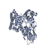
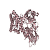
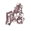
Crystal Structure of Bacillus thuringiensis Cry1A.105 Tryptic Core
Components
Cry1A.105
Keywords
TOXIN / Pesticidal crystal protein
Function / homology
Function and homology information
symbiont-mediated killing of host cell / sporulation resulting in formation of a cellular spore / toxin activity / signaling receptor binding Similarity search - Function
Pesticidal crystal protein Cry1Aa, domain IV / : / Cry1Ac, domain VII / Pesticidal crystal protein, central domain / Pesticidal crystal protein Cry, domain V / Insecticidal delta-endotoxin CryIA(c) domain 5 / Pesticidal crystal protein, N-terminal domain / Pesticidal crystal protein, central domain / delta endotoxin / Pesticidal crystal protein, central domain superfamily ...Pesticidal crystal protein Cry1Aa, domain IV / : / Cry1Ac, domain VII / Pesticidal crystal protein, central domain / Pesticidal crystal protein Cry, domain V / Insecticidal delta-endotoxin CryIA(c) domain 5 / Pesticidal crystal protein, N-terminal domain / Pesticidal crystal protein, central domain / delta endotoxin / Pesticidal crystal protein, central domain superfamily / Pesticidal crystal protein, C-terminal / delta endotoxin / Pesticidal crystal protein / Pesticidal crystal protein, N-terminal / Pesticidal crystal protein, N-terminal domain superfamily / delta endotoxin, N-terminal domain / Aligned Prism / Vitelline Membrane Outer Layer Protein I, subunit A / Delta-Endotoxin; domain 1 / Galactose-binding domain-like / Galactose-binding-like domain superfamily / Jelly Rolls / Up-down Bundle / Sandwich / Mainly Beta / Mainly Alpha Similarity search - Domain/homology
Mass: 18.015 Da / Num. of mol.: 27 / Source method: isolated from a natural source / Formula: H2O
Sequence details
A perfect matching reference sequence is GenBank accession AGN47973.1
-
Experimental details
-
Experiment
Experiment
Method: X-RAY DIFFRACTION / Number of used crystals: 1
-
Sample preparation
Crystal
Density Matthews: 2.27 Å3/Da / Density % sol: 45.81 % / Description: Rectangular, plate-like
Crystal grow
Temperature: 294 K / Method: vapor diffusion, hanging drop / pH: 6 Details: Crystals were obtained using a precipitant/reservoir solution of 5% (w/v) PEG 4000, 0.1 M sodium citrate buffer, pH 6, and 10% isopropanol. Micro-seeding was required to obtain crystals ...Details: Crystals were obtained using a precipitant/reservoir solution of 5% (w/v) PEG 4000, 0.1 M sodium citrate buffer, pH 6, and 10% isopropanol. Micro-seeding was required to obtain crystals sizable enough for X-ray intensity data collection at the synchrotron. PH range: 6
In the structure databanks used in Yorodumi, some data are registered as the other names, "COVID-19 virus" and "2019-nCoV". Here are the details of the virus and the list of structure data.
Jan 31, 2019. EMDB accession codes are about to change! (news from PDBe EMDB page)
EMDB accession codes are about to change! (news from PDBe EMDB page)
The allocation of 4 digits for EMDB accession codes will soon come to an end. Whilst these codes will remain in use, new EMDB accession codes will include an additional digit and will expand incrementally as the available range of codes is exhausted. The current 4-digit format prefixed with “EMD-” (i.e. EMD-XXXX) will advance to a 5-digit format (i.e. EMD-XXXXX), and so on. It is currently estimated that the 4-digit codes will be depleted around Spring 2019, at which point the 5-digit format will come into force.
The EM Navigator/Yorodumi systems omit the EMD- prefix.
Related info.:Q: What is EMD? / ID/Accession-code notation in Yorodumi/EM Navigator
Yorodumi is a browser for structure data from EMDB, PDB, SASBDB, etc.
This page is also the successor to EM Navigator detail page, and also detail information page/front-end page for Omokage search.
The word "yorodu" (or yorozu) is an old Japanese word meaning "ten thousand". "mi" (miru) is to see.
Related info.:EMDB / PDB / SASBDB / Comparison of 3 databanks / Yorodumi Search / Aug 31, 2016. New EM Navigator & Yorodumi / Yorodumi Papers / Jmol/JSmol / Function and homology information / Changes in new EM Navigator and Yorodumi
 Movie
Movie Controller

 Yorodumi
 Open data
 Basic information
 Components
 Keywords
 Function and homology information
 X-RAY DIFFRACTION /
  Authors
 Citation
 Structure visualization
 Downloads & links
 Other downloads
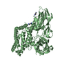


 PDBj
PDBj Assembly
 Mass: 18.015 Da / Num. of mol.: 27 / Source method: isolated from a natural source / Formula: H2O
 Sample preparation
 / Beamline: 22-ID / Wavelength: 1 Å
 Processing