 Movie
Movie Controller


+ Open data

- Basic information
| Entry | Database: PDB / ID: 6dft | ||||||
|---|---|---|---|---|---|---|---|
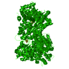
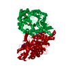
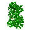
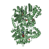
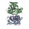
| Title | Trypanosoma brucei deoxyhypusine synthase | ||||||
 Components Components |
| ||||||
 Keywords Keywords | TRANSFERASE / heterotetramer / Rossman fold / pseudoenzyme / NAD+ | ||||||
| Function / homology |  Function and homology information Function and homology informationpeptidyl-hypusine biosynthetic process / regulation of cytoplasmic translation / deoxyhypusine synthase / positive regulation of catalytic activity / deoxyhypusine synthase activity / spermidine metabolic process / protein heterotetramerization / catalytic complex / enzyme activator activity / enzyme binding ...peptidyl-hypusine biosynthetic process / regulation of cytoplasmic translation / deoxyhypusine synthase / positive regulation of catalytic activity / deoxyhypusine synthase activity / spermidine metabolic process / protein heterotetramerization / catalytic complex / enzyme activator activity / enzyme binding / protein homodimerization activity / membrane / cytoplasm Similarity search - Function | ||||||
| Biological species |  | ||||||
| Method |  X-RAY DIFFRACTION / SYNCHROTRON / MOLECULAR REPLACEMENT / Resolution: 3.5 Å X-RAY DIFFRACTION / SYNCHROTRON / MOLECULAR REPLACEMENT / Resolution: 3.5 Å | ||||||
| Model details | Active enzyme is a heterotetramer, composed of a heterodimer of the catalytic and pseudoenzyme monomers. | ||||||
 Authors Authors | Tomchick, D.R. / Phillips, M.A. / Afanador, G.A. | ||||||
| Funding support |  United States, 1items United States, 1items
| ||||||
 Citation Citation | Journal: Structure / Year: 2018 Title: Trypanosomatid Deoxyhypusine Synthase Activity Is Dependent on Shared Active-Site Complementation between Pseudoenzyme Paralogs. Authors: Afanador, G.A. / Tomchick, D.R. / Phillips, M.A. #1: Journal: J Biol Chem / Year: 2013 Title: Allosteric Activation of Trypanosomatid Deoxyhypusine Synthase by a Catalytically Dead Paralog Authors: Suong, N. / Jones, D.C. / Wyllie, S. / Fairlamb, A.H. / Phillips, M.A. | ||||||
| History |
|
- Structure visualization
| Structure viewer | Molecule: MolmilJmol/JSmol |
|---|
- Downloads & links
-Download
| PDBx/mmCIF format | 6dft.cif.gz | 786.5 KB | Display | PDBx/mmCIF format |
|---|---|---|---|---|
| PDB format | pdb6dft.ent.gz | 653.9 KB | Display | PDB format |
| PDBx/mmJSON format | 6dft.json.gz | Tree view | PDBx/mmJSON format | |
| Others |  Other downloads Other downloads |
-Validation report
| Arichive directory | https://data.pdbj.org/pub/pdb/validation_reports/df/6dftftp://data.pdbj.org/pub/pdb/validation_reports/df/6dft | HTTPS FTP |
|---|
-Related structure data
| Related structure data |  1rqdS S: Starting model for refinement |
|---|---|
| Similar structure data |


-Links
 PDBj
PDBj

- Assembly
| Deposited unit | 
| ||||||||
|---|---|---|---|---|---|---|---|---|---|
| 1 | 
| ||||||||
| 2 | 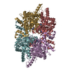
| ||||||||
| 3 | 
| ||||||||
| Unit cell |
|
-Components
| #1: Protein | Mass: 50226.137 Da / Num. of mol.: 6 / Fragment: catalytic monomer Source method: isolated from a genetically manipulated source Source: (gene. exp.)  #2: Protein | Mass: 37419.312 Da / Num. of mol.: 6 / Fragment: pseudoenzyme monomer Source method: isolated from a genetically manipulated source Source: (gene. exp.) #3: Chemical | ChemComp-NA / |   Mass: 22.990 Da / Num. of mol.: 1 / Source method: obtained synthetically / Formula: Na Mass: 22.990 Da / Num. of mol.: 1 / Source method: obtained synthetically / Formula: Na#4: Chemical | ChemComp-NAD /   Mass: 663.425 Da / Num. of mol.: 12 / Source method: obtained synthetically / Formula: C21H27N7O14P2 / Feature type: SUBJECT OF INVESTIGATION / Comment: NAD*YM Mass: 663.425 Da / Num. of mol.: 12 / Source method: obtained synthetically / Formula: C21H27N7O14P2 / Feature type: SUBJECT OF INVESTIGATION / Comment: NAD*YM |
|---|
-Experimental details
-Experiment
| Experiment | Method: X-RAY DIFFRACTION / Number of used crystals: 1 |
|---|
- Sample preparation
| Crystal | Density Matthews: 1.98 Å3/Da / Density % sol: 37.66 % / Description: THIN PLATE |
|---|---|
| Crystal grow | Temperature: 293 K / Method: vapor diffusion, sitting drop / pH: 7 Details: 0.1 M HEPES, 10 mM TCEP, 2 mM NAD+, 8% PEG 6000, 20% ETHYLENE GLYCOL |
-Data collection
| Diffraction | Mean temperature: 100 K | |||||||||||||||||||||||||||||||||||||||||||||||||||||||||||||||||||||||||||||||||||||||||||||||||||||||||||||||||||||||||||||||||||||||||||||||||||||||||||||||||||||||||||||||||||||||||||||
|---|---|---|---|---|---|---|---|---|---|---|---|---|---|---|---|---|---|---|---|---|---|---|---|---|---|---|---|---|---|---|---|---|---|---|---|---|---|---|---|---|---|---|---|---|---|---|---|---|---|---|---|---|---|---|---|---|---|---|---|---|---|---|---|---|---|---|---|---|---|---|---|---|---|---|---|---|---|---|---|---|---|---|---|---|---|---|---|---|---|---|---|---|---|---|---|---|---|---|---|---|---|---|---|---|---|---|---|---|---|---|---|---|---|---|---|---|---|---|---|---|---|---|---|---|---|---|---|---|---|---|---|---|---|---|---|---|---|---|---|---|---|---|---|---|---|---|---|---|---|---|---|---|---|---|---|---|---|---|---|---|---|---|---|---|---|---|---|---|---|---|---|---|---|---|---|---|---|---|---|---|---|---|---|---|---|---|---|---|---|---|
| Diffraction source | Source: SYNCHROTRON / Site: APS / Beamline: 19-ID / Wavelength: 0.97918 Å | |||||||||||||||||||||||||||||||||||||||||||||||||||||||||||||||||||||||||||||||||||||||||||||||||||||||||||||||||||||||||||||||||||||||||||||||||||||||||||||||||||||||||||||||||||||||||||||
| Detector | Type: DECTRIS PILATUS3 6M / Detector: PIXEL / Date: Sep 20, 2017 / Details: monochromator | |||||||||||||||||||||||||||||||||||||||||||||||||||||||||||||||||||||||||||||||||||||||||||||||||||||||||||||||||||||||||||||||||||||||||||||||||||||||||||||||||||||||||||||||||||||||||||||
| Radiation | Protocol: SINGLE WAVELENGTH / Monochromatic (M) / Laue (L): M / Scattering type: x-ray | |||||||||||||||||||||||||||||||||||||||||||||||||||||||||||||||||||||||||||||||||||||||||||||||||||||||||||||||||||||||||||||||||||||||||||||||||||||||||||||||||||||||||||||||||||||||||||||
| Radiation wavelength | Wavelength: 0.97918 Å / Relative weight: 1 | |||||||||||||||||||||||||||||||||||||||||||||||||||||||||||||||||||||||||||||||||||||||||||||||||||||||||||||||||||||||||||||||||||||||||||||||||||||||||||||||||||||||||||||||||||||||||||||
| Reflection | Resolution: 3.5→50 Å / Num. obs: 52848 / % possible obs: 97.2 % / Redundancy: 4.1 % / Biso Wilson estimate: 64.48 Å2 / Rmerge(I) obs: 0.188 / Rpim(I) all: 0.101 / Rrim(I) all: 0.214 / Χ2: 0.87 / Net I/σ(I): 4.1 / Num. measured all: 214291 | |||||||||||||||||||||||||||||||||||||||||||||||||||||||||||||||||||||||||||||||||||||||||||||||||||||||||||||||||||||||||||||||||||||||||||||||||||||||||||||||||||||||||||||||||||||||||||||
| Reflection shell | Diffraction-ID: 1
|
- Processing
| Software |
| |||||||||||||||||||||||||||||||||||||||||||||||||||||||||||||||||||||||||||||||||||||||||||||||||||||||||
|---|---|---|---|---|---|---|---|---|---|---|---|---|---|---|---|---|---|---|---|---|---|---|---|---|---|---|---|---|---|---|---|---|---|---|---|---|---|---|---|---|---|---|---|---|---|---|---|---|---|---|---|---|---|---|---|---|---|---|---|---|---|---|---|---|---|---|---|---|---|---|---|---|---|---|---|---|---|---|---|---|---|---|---|---|---|---|---|---|---|---|---|---|---|---|---|---|---|---|---|---|---|---|---|---|---|---|
| Refinement | Method to determine structure: MOLECULAR REPLACEMENT Starting model: 1RQD Resolution: 3.5→48.752 Å / SU ML: 0.46 / Cross valid method: FREE R-VALUE / σ(F): 1.35 / Phase error: 26.62
| |||||||||||||||||||||||||||||||||||||||||||||||||||||||||||||||||||||||||||||||||||||||||||||||||||||||||
| Solvent computation | Shrinkage radii: 0.9 Å / VDW probe radii: 1.11 Å | |||||||||||||||||||||||||||||||||||||||||||||||||||||||||||||||||||||||||||||||||||||||||||||||||||||||||
| Displacement parameters | Biso max: 147.42 Å2 / Biso mean: 57.1506 Å2 / Biso min: 23.31 Å2 | |||||||||||||||||||||||||||||||||||||||||||||||||||||||||||||||||||||||||||||||||||||||||||||||||||||||||
| Refinement step | Cycle: final / Resolution: 3.5→48.752 Å
| |||||||||||||||||||||||||||||||||||||||||||||||||||||||||||||||||||||||||||||||||||||||||||||||||||||||||
| LS refinement shell | Refine-ID: X-RAY DIFFRACTION / Rfactor Rfree error: 0 / Total num. of bins used: 14
|
