 Movie
Movie Controller


+ Open data

- Basic information
| Entry | Database: PDB / ID: 6d00 | |||||||||||||||||||||
|---|---|---|---|---|---|---|---|---|---|---|---|---|---|---|---|---|---|---|---|---|---|---|
| Title | Calcarisporiella thermophila Hsp104 | |||||||||||||||||||||
 Components Components | Calcarisporiella thermophila Hsp104 | |||||||||||||||||||||
 Keywords Keywords | CHAPERONE / Spiral hexamer / antagonize toxin / ADP-binding state | |||||||||||||||||||||
| Function / homology |  Function and homology information Function and homology informationcellular heat acclimation / protein unfolding / : / protein refolding / protein-folding chaperone binding / ATP hydrolysis activity / ATP binding / cytosol Similarity search - Function | |||||||||||||||||||||
| Biological species |  Calcarisporiella thermophila (fungus) Calcarisporiella thermophila (fungus) | |||||||||||||||||||||
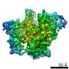
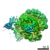
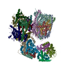
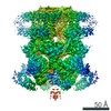
| Method | ELECTRON MICROSCOPY / single particle reconstruction / cryo EM / Resolution: 4 Å | |||||||||||||||||||||
 Authors Authors | Zhang, K. / Pintilie, G. | |||||||||||||||||||||
| Funding support |  United States, 6items United States, 6items
| |||||||||||||||||||||
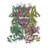
 Citation Citation | Journal: Structure / Year: 2019 Title: Structure of Calcarisporiella thermophila Hsp104 Disaggregase that Antagonizes Diverse Proteotoxic Misfolding Events. Authors: Karolina Michalska / Kaiming Zhang / Zachary M March / Catherine Hatzos-Skintges / Grigore Pintilie / Lance Bigelow / Laura M Castellano / Leann J Miles / Meredith E Jackrel / Edward Chuang ...Authors: Karolina Michalska / Kaiming Zhang / Zachary M March / Catherine Hatzos-Skintges / Grigore Pintilie / Lance Bigelow / Laura M Castellano / Leann J Miles / Meredith E Jackrel / Edward Chuang / Robert Jedrzejczak / James Shorter / Wah Chiu / Andrzej Joachimiak / Abstract: Hsp104 is an AAA+ protein disaggregase with powerful amyloid-remodeling activity. All nonmetazoan eukaryotes express Hsp104 while eubacteria express an Hsp104 ortholog, ClpB. However, most studies ...Hsp104 is an AAA+ protein disaggregase with powerful amyloid-remodeling activity. All nonmetazoan eukaryotes express Hsp104 while eubacteria express an Hsp104 ortholog, ClpB. However, most studies have focused on Hsp104 from Saccharomyces cerevisiae and ClpB orthologs from two eubacterial species. Thus, the natural spectrum of Hsp104/ClpB molecular architectures and protein-remodeling activities remains largely unexplored. Here, we report two structures of Hsp104 from the thermophilic fungus Calcarisporiella thermophila (CtHsp104), a 2.70Å crystal structure and 4.0Å cryo-electron microscopy structure. Both structures reveal left-handed, helical assemblies with all domains clearly resolved. We thus provide the highest resolution and most complete view of Hsp104 hexamers to date. We also establish that CtHsp104 antagonizes several toxic protein-misfolding events in vivo where S. cerevisiae Hsp104 is ineffective, including rescue of TDP-43, polyglutamine, and α-synuclein toxicity. We suggest that natural Hsp104 variation is an invaluable, untapped resource for illuminating therapeutic disaggregases for fatal neurodegenerative diseases. | |||||||||||||||||||||
| History |
|
- Structure visualization
| Movie |
Movie viewer |
|---|---|
| Structure viewer | Molecule: MolmilJmol/JSmol |
- Downloads & links
-Download
| PDBx/mmCIF format | 6d00.cif.gz | 893.3 KB | Display | PDBx/mmCIF format |
|---|---|---|---|---|
| PDB format | pdb6d00.ent.gz | 723.7 KB | Display | PDB format |
| PDBx/mmJSON format | 6d00.json.gz | Tree view | PDBx/mmJSON format | |
| Others |  Other downloads Other downloads |
-Validation report
| Arichive directory | https://data.pdbj.org/pub/pdb/validation_reports/d0/6d00ftp://data.pdbj.org/pub/pdb/validation_reports/d0/6d00 | HTTPS FTP |
|---|
-Related structure data
| Related structure data |  7782MC  6azyC C: citing same article ( M: map data used to model this data |
|---|---|
| Similar structure data |


-Links
 PDBj
PDBj


- Assembly
| Deposited unit | 
|
|---|---|
| 1 |
|
-Components
| #1: Protein | Mass: 98911.109 Da / Num. of mol.: 6 Source method: isolated from a genetically manipulated source Source: (gene. exp.) Calcarisporiella thermophila (fungus)Production host:  References: UniProt: A0A452CSQ7*PLUS #2: Chemical | ChemComp-ADP /   Mass: 427.201 Da / Num. of mol.: 12 / Source method: obtained synthetically / Formula: C10H15N5O10P2 / Comment: ADP, energy-carrying molecule*YM Mass: 427.201 Da / Num. of mol.: 12 / Source method: obtained synthetically / Formula: C10H15N5O10P2 / Comment: ADP, energy-carrying molecule*YM |
|---|
-Experimental details
-Experiment
| Experiment | Method: ELECTRON MICROSCOPY |
|---|---|
| EM experiment | Aggregation state: PARTICLE / 3D reconstruction method: single particle reconstruction |
- Sample preparation
| Component | Name: Calcarisporiella thermophila Hsp104 with ADP / Type: COMPLEX / Entity ID: #1 / Source: RECOMBINANT |
|---|---|
| Molecular weight | Value: 0.6 MDa / Experimental value: YES |
| Source (natural) | Organism: Calcarisporiella thermophila (fungus) |
| Source (recombinant) | Organism: |
| Buffer solution | pH: 8 Details: 20 mM HEPES-KOH, 100 mM KCl, 2 mM MgCl2, 2 mM ADP and 4 mM DTT |
| Specimen | Conc.: 0.5 mg/ml / Embedding applied: NO / Shadowing applied: NO / Staining applied: NO / Vitrification applied: YES |
| Specimen support | Grid material: COPPER / Grid mesh size: 200 divisions/in. / Grid type: Quantifoil R2/1 |
| Vitrification | Instrument: FEI VITROBOT MARK IV / Cryogen name: ETHANE / Humidity: 100 % |
- Electron microscopy imaging
| Experimental equipment |  Model: Titan Krios / Image courtesy: FEI Company |
|---|---|
| Microscopy | Model: FEI TITAN KRIOS |
| Electron gun | Electron source:  FIELD EMISSION GUN / Accelerating voltage: 300 kV / Illumination mode: FLOOD BEAM FIELD EMISSION GUN / Accelerating voltage: 300 kV / Illumination mode: FLOOD BEAM |
| Electron lens | Mode: BRIGHT FIELD / Nominal magnification: 130000 X / Nominal defocus max: 2000 nm / Nominal defocus min: 1000 nm / Calibrated defocus min: 800 nm / Calibrated defocus max: 2500 nm / Cs: 2.7 mm / C2 aperture diameter: 70 µm / Alignment procedure: BASIC |
| Specimen holder | Cryogen: NITROGEN / Specimen holder model: FEI TITAN KRIOS AUTOGRID HOLDER |
| Image recording | Average exposure time: 6 sec. / Electron dose: 31.8 e/Å2 / Detector mode: COUNTING / Film or detector model: GATAN K2 SUMMIT (4k x 4k) / Num. of grids imaged: 2 / Num. of real images: 3786 |
| EM imaging optics | Energyfilter name: GIF Quantum LS |
| Image scans | Movie frames/image: 30 / Used frames/image: 3-30 |
- Processing
| Software |
| ||||||||||||||||||||
|---|---|---|---|---|---|---|---|---|---|---|---|---|---|---|---|---|---|---|---|---|---|
| EM software |
| ||||||||||||||||||||
| CTF correction | Details: Gctf / Type: PHASE FLIPPING AND AMPLITUDE CORRECTION | ||||||||||||||||||||
| Particle selection | Num. of particles selected: 372498 | ||||||||||||||||||||
| Symmetry | Point symmetry: C1 (asymmetric) | ||||||||||||||||||||
| 3D reconstruction | Resolution: 4 Å / Resolution method: FSC 0.143 CUT-OFF / Num. of particles: 224915 / Symmetry type: POINT | ||||||||||||||||||||
| Atomic model building | Protocol: FLEXIBLE FIT |
