[English] 日本語
Yorodumi
- EMDB-7782: Calcarisporiella thermophila Hsp104 -

+
Open data


ID or keywords:

Loading...

-
Basic information

Entry
Database: EMDB / ID: EMD-7782
TitleCalcarisporiella thermophila Hsp104
Map dataMap was masked with model and equalized to bring out some of the more flexible domains.
Sample
  • Complex: Calcarisporiella thermophila Hsp104 with ADP
    • Protein or peptide: Calcarisporiella thermophila Hsp104
  • Ligand: ADENOSINE-5'-DIPHOSPHATE
KeywordsSpiral hexamer / antagonize toxin / ADP-binding state / CHAPERONE
Function / homology
Function and homology information


ATP hydrolysis activity / ATP binding
Similarity search - Function
ClpA/B, conserved site 2 / Chaperonins clpA/B signature 2. / ClpA/B, conserved site 1 / Chaperonins clpA/B signature 1. / ClpA/ClpB, AAA lid domain / AAA lid domain / Clp amino terminal domain, pathogenicity island component / Clp repeat (R) domain profile. / Clp, repeat (R) domain / Clp, N-terminal domain superfamily ...ClpA/B, conserved site 2 / Chaperonins clpA/B signature 2. / ClpA/B, conserved site 1 / Chaperonins clpA/B signature 1. / ClpA/ClpB, AAA lid domain / AAA lid domain / Clp amino terminal domain, pathogenicity island component / Clp repeat (R) domain profile. / Clp, repeat (R) domain / Clp, N-terminal domain superfamily / ClpA/B family / Clp ATPase, C-terminal / AAA domain (Cdc48 subfamily) / C-terminal, D2-small domain, of ClpB protein / C-terminal, D2-small domain, of ClpB protein / ATPase family associated with various cellular activities (AAA) / ATPase, AAA-type, core / ATPases associated with a variety of cellular activities / AAA+ ATPase domain / P-loop containing nucleoside triphosphate hydrolase
Similarity search - Domain/homology
Heat shock protein Hsp104
Similarity search - Component
Biological speciesCalcarisporiella thermophila (fungus)
Methodsingle particle reconstruction / cryo EM / Resolution: 4.0 Å
AuthorsZhang K / Pintilie G
Funding support United States, 6 items
OrganizationGrant numberCountry
National Institutes of Health/National Human Genome Research Institute (NIH/NHGRI)T32GM071399 United States
National Institutes of Health/National Human Genome Research Institute (NIH/NHGRI)F31NS101807 United States
National Institutes of Health/National Human Genome Research Institute (NIH/NHGRI)R01GM099836 United States
Department of Energy (DOE, United States)GM094585 United States
Department of Energy (DOE, United States)GM115586 United States
National Institutes of Health/National Human Genome Research Institute (NIH/NHGRI)P41GM103832 United States
Citation
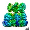
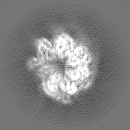
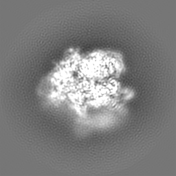
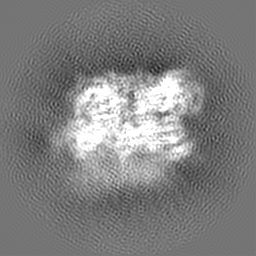
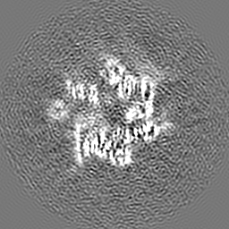
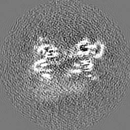
Journal: Structure / Year: 2019
Title: Structure of Calcarisporiella thermophila Hsp104 Disaggregase that Antagonizes Diverse Proteotoxic Misfolding Events.
Authors: Karolina Michalska / Kaiming Zhang / Zachary M March / Catherine Hatzos-Skintges / Grigore Pintilie / Lance Bigelow / Laura M Castellano / Leann J Miles / Meredith E Jackrel / Edward Chuang ...Authors: Karolina Michalska / Kaiming Zhang / Zachary M March / Catherine Hatzos-Skintges / Grigore Pintilie / Lance Bigelow / Laura M Castellano / Leann J Miles / Meredith E Jackrel / Edward Chuang / Robert Jedrzejczak / James Shorter / Wah Chiu / Andrzej Joachimiak /
Abstract: Hsp104 is an AAA+ protein disaggregase with powerful amyloid-remodeling activity. All nonmetazoan eukaryotes express Hsp104 while eubacteria express an Hsp104 ortholog, ClpB. However, most studies ...Hsp104 is an AAA+ protein disaggregase with powerful amyloid-remodeling activity. All nonmetazoan eukaryotes express Hsp104 while eubacteria express an Hsp104 ortholog, ClpB. However, most studies have focused on Hsp104 from Saccharomyces cerevisiae and ClpB orthologs from two eubacterial species. Thus, the natural spectrum of Hsp104/ClpB molecular architectures and protein-remodeling activities remains largely unexplored. Here, we report two structures of Hsp104 from the thermophilic fungus Calcarisporiella thermophila (CtHsp104), a 2.70Å crystal structure and 4.0Å cryo-electron microscopy structure. Both structures reveal left-handed, helical assemblies with all domains clearly resolved. We thus provide the highest resolution and most complete view of Hsp104 hexamers to date. We also establish that CtHsp104 antagonizes several toxic protein-misfolding events in vivo where S. cerevisiae Hsp104 is ineffective, including rescue of TDP-43, polyglutamine, and α-synuclein toxicity. We suggest that natural Hsp104 variation is an invaluable, untapped resource for illuminating therapeutic disaggregases for fatal neurodegenerative diseases.
History
DepositionApr 9, 2018-
Header (metadata) releaseJul 11, 2018-
Map releaseApr 3, 2019-
UpdateMar 13, 2024-
Current statusMar 13, 2024Processing site: RCSB / Status: Released

-
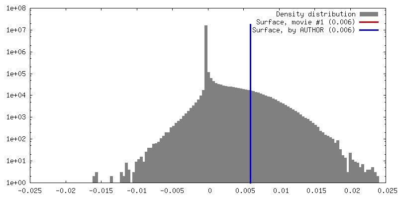
Structure visualization
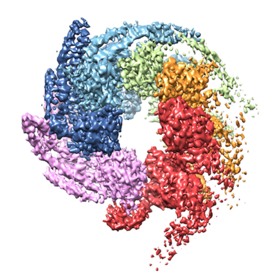
Movie
  • Surface view with section colored by density value
  • Surface level: 0.006
  • Imaged by UCSF Chimera
  • Download
  • Surface view colored by cylindrical radius
  • Surface level: 0.006
  • Imaged by UCSF Chimera
  • Download
  • Surface view with fitted model
  • Atomic models: PDB-6d00
  • Surface level: 0.006
  • Imaged by UCSF Chimera
  • Download
Movie viewer
Structure viewerEM map:
SurfViewMolmilJmol/JSmol
Supplemental images

Downloads & links

-
Map

FileDownload / File: emd_7782.map.gz / Format: CCP4 / Size: 64 MB / Type: IMAGE STORED AS FLOATING POINT NUMBER (4 BYTES)
AnnotationMap was masked with model and equalized to bring out some of the more flexible domains.
Voxel sizeX=Y=Z: 1.06 Å
Density
Contour LevelBy AUTHOR: 0.006 / Movie #1: 0.006
Minimum - Maximum-0.019896325 - 0.024561299
Average (Standard dev.)0.0001508107 (±0.0009997501)
SymmetrySpace group: 1
Details

EMDB XML:

Map geometry
Axis orderXYZ
Origin000
Dimensions256256256
Spacing256256256
CellA=B=C: 271.36 Å
α=β=γ: 90.0 °

CCP4 map header:

modeImage stored as Reals
Å/pix. X/Y/Z1.061.061.06
M x/y/z256256256
origin x/y/z0.0000.0000.000
length x/y/z271.360271.360271.360
α/β/γ90.00090.00090.000
start NX/NY/NZ-383-383-383
NX/NY/NZ768768768
MAP C/R/S123
start NC/NR/NS000
NC/NR/NS256256256
D min/max/mean-0.0200.0250.000

-
Supplemental data

-
Additional map: Original reconstructed map.

Fileemd_7782_additional.map
AnnotationOriginal reconstructed map.
Projections & Slices
AxesZYX

Projections

Slices (1/2)
Density Histograms

-
Sample components

-
Entire : Calcarisporiella thermophila Hsp104 with ADP

EntireName: Calcarisporiella thermophila Hsp104 with ADP
Components
  • Complex: Calcarisporiella thermophila Hsp104 with ADP
    • Protein or peptide: Calcarisporiella thermophila Hsp104
  • Ligand: ADENOSINE-5'-DIPHOSPHATE

-
Supramolecule #1: Calcarisporiella thermophila Hsp104 with ADP

SupramoleculeName: Calcarisporiella thermophila Hsp104 with ADP / type: complex / ID: 1 / Parent: 0 / Macromolecule list: #1
Source (natural)Organism: Calcarisporiella thermophila (fungus)
Molecular weightTheoretical: 600 KDa

-
Macromolecule #1: Calcarisporiella thermophila Hsp104

MacromoleculeName: Calcarisporiella thermophila Hsp104 / type: protein_or_peptide / ID: 1 / Number of copies: 6 / Enantiomer: LEVO
Source (natural)Organism: Calcarisporiella thermophila (fungus)
Molecular weightTheoretical: 98.911109 KDa
Recombinant expressionOrganism: Escherichia coli 'BL21-Gold(DE3)pLysS AG' (bacteria)
SequenceString: AMSSMQFTDK ATETLNAAAK YAAENSHVQL HPSHVAVVML DEENSLFRSI LEKAGGDVVS IERGFKKIMV RQPSQDPPPT EMGHSPELA KLLHYAHEHM KKQRDLYIAQ DHLILALADL PSMAQVLKEG GVTKKSLENA VTHVRGNRRV ESKSAEEAYE A LSKYCIDL ...String:
AMSSMQFTDK ATETLNAAAK YAAENSHVQL HPSHVAVVML DEENSLFRSI LEKAGGDVVS IERGFKKIMV RQPSQDPPPT EMGHSPELA KLLHYAHEHM KKQRDLYIAQ DHLILALADL PSMAQVLKEG GVTKKSLENA VTHVRGNRRV ESKSAEEAYE A LSKYCIDL TELAASGKLD PVIGRDEIIS RVIRVLSRRT KNNPCLVGEP GVGKTAIAEG LANRIVKGDI PSSLQKKVYS LD IGSLLAG AKYRGEFEER LKAVLKELKE AQAIVFIDEI HTVLGAGKSE GAIDAANLLK PMLARGELRC IGATTLTEYR QYV EKDPAF ERRFQLVMVE EPSVTDTISI LRGLKERYET HHGVRIADAA IVAAAQLAAR YITQRFMPDK AIDLIDEACA NTRV QLDSQ PEAIDKLERR HLQLEVEATA LEKEKDAASK QRLQEVRAEM ARIQEELRPL KMKYESEKGR LDEIRNLSQR LDELK AKAE DAERRYDLAR AADIRYYAIP DLEKRLAQLQ AEKSQADAER ADGLLAEVVG PDQIMEVVSR WTGIPVSNLQ RSEKEK LLH MEEYMKQHVV GQDEAIKAIC DAIRLSRTGL QNRNRPLASF LFLGPTGCGK TLCVKELAAF LFNDPGAIVR IDMSEYM EK HAVSRLVGAP PGYIGHDEGG QLTEAVRRRP YTVVLFDEME KAHKDVSNLL LQILDDGHCT DSKGRRVDFK NTIIVMTS N LGADLFELDE GDKVSQATKN AVLATARRHF ANEFINRIDE LIVFNRLTPS NIRKIVDVRL KEVQERLDEK QITLDVDDK AKDLLAQQGF DPVYGARPLN RLIQHALLTQ LSRLLLDGGV RPGEIAKVTV DQEGEIIVIR NHGIESPAPW ADEDMVEDED MEI

-
Macromolecule #2: ADENOSINE-5'-DIPHOSPHATE

MacromoleculeName: ADENOSINE-5'-DIPHOSPHATE / type: ligand / ID: 2 / Number of copies: 12 / Formula: ADP
Molecular weightTheoretical: 427.201 Da
Chemical component information

ChemComp-ADP:
ADENOSINE-5'-DIPHOSPHATE / ADP, energy-carrying molecule*YM / Adenosine diphosphate

-
Experimental details

-
Structure determination

Methodcryo EM
Processingsingle particle reconstruction
Aggregation stateparticle

-
Sample preparation

Concentration0.5 mg/mL
BufferpH: 8
Details: 20 mM HEPES-KOH, 100 mM KCl, 2 mM MgCl2, 2 mM ADP and 4 mM DTT
GridModel: Quantifoil R2/1 / Material: COPPER / Mesh: 200 / Pretreatment - Type: GLOW DISCHARGE / Pretreatment - Time: 25 sec.
VitrificationCryogen name: ETHANE / Chamber humidity: 100 % / Instrument: FEI VITROBOT MARK IV

-
Electron microscopy

MicroscopeFEI TITAN KRIOS
Electron beamAcceleration voltage: 300 kV / Electron source: FIELD EMISSION GUN
Electron opticsC2 aperture diameter: 70.0 µm / Calibrated defocus max: 2.5 µm / Calibrated defocus min: 0.8 µm / Illumination mode: FLOOD BEAM / Imaging mode: BRIGHT FIELDBright-field microscopy / Cs: 2.7 mm / Nominal defocus max: 2.0 µm / Nominal defocus min: 1.0 µm / Nominal magnification: 130000
Specialist opticsEnergy filter - Name: GIF Quantum LS
Sample stageSpecimen holder model: FEI TITAN KRIOS AUTOGRID HOLDER / Cooling holder cryogen: NITROGEN
Image recordingFilm or detector model: GATAN K2 SUMMIT (4k x 4k) / Detector mode: COUNTING / Digitization - Frames/image: 3-30 / Number grids imaged: 2 / Number real images: 3786 / Average exposure time: 6.0 sec. / Average electron dose: 31.8 e/Å2
Experimental equipment
Model: Titan Krios / Image courtesy: FEI Company

-
Image processing

Particle selectionNumber selected: 372498
Startup modelType of model: INSILICO MODEL
Initial angle assignmentType: COMMON LINE
Final angle assignmentType: COMMON LINE
Final reconstructionApplied symmetry - Point group: C1 (asymmetric) / Resolution.type: BY AUTHOR / Resolution: 4.0 Å / Resolution method: FSC 0.143 CUT-OFF / Software - Name: RELION (ver. 2.1) / Number images used: 224915

-
Atomic model buiding 1

RefinementProtocol: FLEXIBLE FIT
Output model

PDB-6d00:
Calcarisporiella thermophila Hsp104

+
About Yorodumi

-
News

-
Feb 9, 2022. New format data for meta-information of EMDB entries

New format data for meta-information of EMDB entries

  • Version 3 of the EMDB header file is now the official format.
  • The previous official version 1.9 will be removed from the archive.

Related info.:EMDB header

External links:wwPDB to switch to version 3 of the EMDB data model

-
Aug 12, 2020. Covid-19 info

Covid-19 info

URL: https://pdbj.org/emnavi/covid19.php

New page: Covid-19 featured information page in EM Navigator.

Related info.:Covid-19 info / Mar 5, 2020. Novel coronavirus structure data

+
Mar 5, 2020. Novel coronavirus structure data

Novel coronavirus structure data

Related info.:Yorodumi Speices / Aug 12, 2020. Covid-19 info

External links:COVID-19 featured content - PDBj / Molecule of the Month (242):Coronavirus Proteases

+
Jan 31, 2019. EMDB accession codes are about to change! (news from PDBe EMDB page)

EMDB accession codes are about to change! (news from PDBe EMDB page)

  • The allocation of 4 digits for EMDB accession codes will soon come to an end. Whilst these codes will remain in use, new EMDB accession codes will include an additional digit and will expand incrementally as the available range of codes is exhausted. The current 4-digit format prefixed with “EMD-” (i.e. EMD-XXXX) will advance to a 5-digit format (i.e. EMD-XXXXX), and so on. It is currently estimated that the 4-digit codes will be depleted around Spring 2019, at which point the 5-digit format will come into force.
  • The EM Navigator/Yorodumi systems omit the EMD- prefix.

Related info.:Q: What is EMD? / ID/Accession-code notation in Yorodumi/EM Navigator

External links:EMDB Accession Codes are Changing Soon! / Contact to PDBj

+
Jul 12, 2017. Major update of PDB

Major update of PDB

  • wwPDB released updated PDB data conforming to the new PDBx/mmCIF dictionary.
  • This is a major update changing the version number from 4 to 5, and with Remediation, in which all the entries are updated.
  • In this update, many items about electron microscopy experimental information are reorganized (e.g. em_software).
  • Now, EM Navigator and Yorodumi are based on the updated data.

External links:wwPDB Remediation / Enriched Model Files Conforming to OneDep Data Standards Now Available in the PDB FTP Archive

-
Yorodumi

Thousand views of thousand structures

  • Yorodumi is a browser for structure data from EMDB, PDB, SASBDB, etc.
  • This page is also the successor to EM Navigator detail page, and also detail information page/front-end page for Omokage search.
  • The word "yorodu" (or yorozu) is an old Japanese word meaning "ten thousand". "mi" (miru) is to see.

Related info.:EMDB / PDB / SASBDB / Comparison of 3 databanks / Yorodumi Search / Aug 31, 2016. New EM Navigator & Yorodumi / Yorodumi Papers / Jmol/JSmol / Function and homology information / Changes in new EM Navigator and Yorodumi

Read more