[English] 日本語
Yorodumi
- PDB-6azy: Crystal structure of Hsp104 R328M/R757M mutant from Calcarisporie... -

+
Open data


ID or keywords:

Loading...

-
Basic information

Entry
Database: PDB / ID: 6azy
TitleCrystal structure of Hsp104 R328M/R757M mutant from Calcarisporiella thermophila
ComponentsHeat shock protein Hsp104Heat shock response
KeywordsCHAPERONE / disaggregase / AAA+ ATPase / Structural Genomics / PSI-Biology / Midwest Center for Structural Genomics / MCSG
Function / homology
Function and homology information


ATP hydrolysis activity / ATP binding
Similarity search - Function
ClpA/B, conserved site 2 / Chaperonins clpA/B signature 2. / ClpA/B, conserved site 1 / Chaperonins clpA/B signature 1. / ClpA/ClpB, AAA lid domain / AAA lid domain / Clp amino terminal domain, pathogenicity island component / Clp repeat (R) domain profile. / Clp, repeat (R) domain / Clp, N-terminal domain superfamily ...ClpA/B, conserved site 2 / Chaperonins clpA/B signature 2. / ClpA/B, conserved site 1 / Chaperonins clpA/B signature 1. / ClpA/ClpB, AAA lid domain / AAA lid domain / Clp amino terminal domain, pathogenicity island component / Clp repeat (R) domain profile. / Clp, repeat (R) domain / Clp, N-terminal domain superfamily / ClpA/B family / Clp ATPase, C-terminal / AAA domain (Cdc48 subfamily) / C-terminal, D2-small domain, of ClpB protein / C-terminal, D2-small domain, of ClpB protein / ATPase family associated with various cellular activities (AAA) / ATPase, AAA-type, core / ATPases associated with a variety of cellular activities / AAA+ ATPase domain / P-loop containing nucleoside triphosphate hydrolase
Similarity search - Domain/homology
ADENOSINE-5'-DIPHOSPHATE / Heat shock protein Hsp104
Similarity search - Component
Biological speciesCalcarisporiella thermophila (fungus)
MethodX-RAY DIFFRACTION / SYNCHROTRON / MOLECULAR REPLACEMENT / Resolution: 2.7 Å
AuthorsMichalska, K. / Bigelow, L. / Hatzos-Skintges, C. / Jedrzejczak, R. / Joachimiak, A. / Midwest Center for Structural Genomics (MCSG)
Funding support United States, 2items
OrganizationGrant numberCountry
National Institutes of Health/National Institute of General Medical Sciences (NIH/NIGMS)GM094585 United States
National Institutes of Health/National Institute of General Medical Sciences (NIH/NIGMS)GM115586 United States
Citation
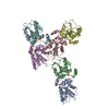
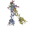
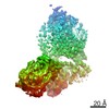
Journal: Structure / Year: 2019
Title: Structure of Calcarisporiella thermophila Hsp104 Disaggregase that Antagonizes Diverse Proteotoxic Misfolding Events.
Authors: Karolina Michalska / Kaiming Zhang / Zachary M March / Catherine Hatzos-Skintges / Grigore Pintilie / Lance Bigelow / Laura M Castellano / Leann J Miles / Meredith E Jackrel / Edward Chuang ...Authors: Karolina Michalska / Kaiming Zhang / Zachary M March / Catherine Hatzos-Skintges / Grigore Pintilie / Lance Bigelow / Laura M Castellano / Leann J Miles / Meredith E Jackrel / Edward Chuang / Robert Jedrzejczak / James Shorter / Wah Chiu / Andrzej Joachimiak /
Abstract: Hsp104 is an AAA+ protein disaggregase with powerful amyloid-remodeling activity. All nonmetazoan eukaryotes express Hsp104 while eubacteria express an Hsp104 ortholog, ClpB. However, most studies ...Hsp104 is an AAA+ protein disaggregase with powerful amyloid-remodeling activity. All nonmetazoan eukaryotes express Hsp104 while eubacteria express an Hsp104 ortholog, ClpB. However, most studies have focused on Hsp104 from Saccharomyces cerevisiae and ClpB orthologs from two eubacterial species. Thus, the natural spectrum of Hsp104/ClpB molecular architectures and protein-remodeling activities remains largely unexplored. Here, we report two structures of Hsp104 from the thermophilic fungus Calcarisporiella thermophila (CtHsp104), a 2.70Å crystal structure and 4.0Å cryo-electron microscopy structure. Both structures reveal left-handed, helical assemblies with all domains clearly resolved. We thus provide the highest resolution and most complete view of Hsp104 hexamers to date. We also establish that CtHsp104 antagonizes several toxic protein-misfolding events in vivo where S. cerevisiae Hsp104 is ineffective, including rescue of TDP-43, polyglutamine, and α-synuclein toxicity. We suggest that natural Hsp104 variation is an invaluable, untapped resource for illuminating therapeutic disaggregases for fatal neurodegenerative diseases.
History
DepositionSep 13, 2017Deposition site: RCSB / Processing site: RCSB
Revision 1.0Oct 3, 2018Provider: repository / Type: Initial release
Revision 1.1Feb 20, 2019Group: Author supporting evidence / Data collection / Category: pdbx_audit_support / Item: _pdbx_audit_support.funding_organization
Revision 1.2Jan 1, 2020Group: Author supporting evidence / Category: pdbx_audit_support / Item: _pdbx_audit_support.funding_organization
Revision 1.3Jul 14, 2021Group: Database references / Category: citation / citation_author
Item: _citation.country / _citation.journal_abbrev ..._citation.country / _citation.journal_abbrev / _citation.journal_id_ASTM / _citation.journal_id_CSD / _citation.journal_id_ISSN / _citation.journal_volume / _citation.page_first / _citation.page_last / _citation.pdbx_database_id_DOI / _citation.pdbx_database_id_PubMed / _citation.title / _citation.year
Revision 1.4Oct 4, 2023Group: Data collection / Database references / Refinement description
Category: chem_comp_atom / chem_comp_bond ...chem_comp_atom / chem_comp_bond / database_2 / pdbx_initial_refinement_model
Item: _database_2.pdbx_DOI / _database_2.pdbx_database_accession

-
Structure visualization

Structure viewerMolecule:
MolmilJmol/JSmol

Downloads & links

-
Assembly

Deposited unit
A: Heat shock protein Hsp104
hetero molecules


Theoretical massNumber of molelcules
Total (without water)99,9153
Polymers99,0601
Non-polymers8542
Water0
1


  • Idetical with deposited unit
  • defined by author&software
  • Evidence: gel filtration
TypeNameSymmetry operationNumber
identity operation1_555x,y,z1
MethodPISA
Unit cell
Length a, b, c (Å)139.173, 139.173, 97.060
Angle α, β, γ (deg.)90.00, 90.00, 120.00
Int Tables number170
Space group name H-MP65
DetailsMonomer as determined by gel filtration

-
Components

#1: Protein Heat shock protein Hsp104 / Heat shock response


Mass: 99060.297 Da / Num. of mol.: 1 / Mutation: R328M, R757M
Source method: isolated from a genetically manipulated source
Source: (gene. exp.) Calcarisporiella thermophila (fungus) / Strain: CBS 279.70 / Gene: Calth2p4_003362 / Plasmid: pMCSG68 / Production host: Escherichia coli (E. coli) / Strain (production host): BL21-Gold (DE3) / References: UniProt: A0A452CSQ7*PLUS
#2: Chemical ChemComp-ADP / ADENOSINE-5'-DIPHOSPHATE / Adenosine diphosphate


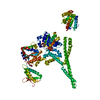
Mass: 427.201 Da / Num. of mol.: 2 / Source method: obtained synthetically / Formula: C10H15N5O10P2 / Comment: ADP, energy-carrying molecule*YM

-
Experimental details

-
Experiment

ExperimentMethod: X-RAY DIFFRACTION / Number of used crystals: 1

-
Sample preparation

CrystalDensity Matthews: 2.74 Å3/Da / Density % sol: 55.1 %
Crystal growTemperature: 277 K / Method: vapor diffusion, sitting drop / pH: 6
Details: 0.2 M Ca acetate, 0.1 M MES/NaOH, pH 6.0, 10% propanol, 5mM ADP, 10 mM MgCl2, cryo 35% glycerol

-
Data collection

DiffractionMean temperature: 100 K
Diffraction sourceSource: SYNCHROTRON / Site: APS / Beamline: 19-ID / Wavelength: 0.9793 Å
DetectorType: ADSC QUANTUM 315r / Detector: CCD / Date: Nov 29, 2014 / Details: mirrors
RadiationMonochromator: Si(111) / Protocol: SINGLE WAVELENGTH / Monochromatic (M) / Laue (L): M / Scattering type: x-ray
Radiation wavelengthWavelength: 0.9793 Å / Relative weight: 1
ReflectionResolution: 2.7→30 Å / Num. obs: 29450 / % possible obs: 100 % / Observed criterion σ(I): -3 / Redundancy: 7.3 % / Biso Wilson estimate: 57.18 Å2 / Rmerge(I) obs: 0.096 / Net I/σ(I): 23.6
Reflection shellResolution: 2.7→2.75 Å / Redundancy: 7.1 % / Rmerge(I) obs: 0 / Mean I/σ(I) obs: 2.25 / Num. unique obs: 1467 / CC1/2: 0.794 / % possible all: 100

-
Processing

Software
NameVersionClassification
PHENIX(dev_2276)refinement
HKL-3000data reduction
HKL-3000data scaling
PHASERphasing
RefinementMethod to determine structure: MOLECULAR REPLACEMENT
Starting model: 1QVR
Resolution: 2.7→29.337 Å / SU ML: 0.37 / Cross valid method: THROUGHOUT / σ(F): 1.35 / Phase error: 27.07
RfactorNum. reflection% reflectionSelection details
Rfree0.2492 1496 5.09 %random
Rwork0.1979 ---
obs0.2005 29413 99.89 %-
Solvent computationShrinkage radii: 0.9 Å / VDW probe radii: 1.11 Å / Solvent model: FLAT BULK SOLVENT MODEL
Refinement stepCycle: LAST / Resolution: 2.7→29.337 Å
ProteinNucleic acidLigandSolventTotal
Num. atoms6372 0 54 0 6426
Refine LS restraints
Refine-IDTypeDev idealNumber
X-RAY DIFFRACTIONf_bond_d0.0086531
X-RAY DIFFRACTIONf_angle_d1.1678805
X-RAY DIFFRACTIONf_dihedral_angle_d10.1337323
X-RAY DIFFRACTIONf_chiral_restr0.0651014
X-RAY DIFFRACTIONf_plane_restr0.0071167
LS refinement shell
Resolution (Å)Rfactor RfreeNum. reflection RfreeRfactor RworkNum. reflection RworkRefine-ID% reflection obs (%)
2.7003-2.78740.27811250.23132519X-RAY DIFFRACTION99
2.7874-2.88690.30661500.23862505X-RAY DIFFRACTION100
2.8869-3.00240.36451160.25492534X-RAY DIFFRACTION100
3.0024-3.13890.33361290.26432557X-RAY DIFFRACTION100
3.1389-3.30420.33771340.23582524X-RAY DIFFRACTION100
3.3042-3.51090.29491440.21852526X-RAY DIFFRACTION100
3.5109-3.78150.22021470.20542520X-RAY DIFFRACTION100
3.7815-4.1610.21711510.17892514X-RAY DIFFRACTION100
4.161-4.76090.21811430.16512549X-RAY DIFFRACTION100
4.7609-5.98990.24731380.20052553X-RAY DIFFRACTION100
5.9899-29.33920.20491190.16862616X-RAY DIFFRACTION100
Refinement TLS params.

Method: refined / Refine-ID: X-RAY DIFFRACTION

IDL112)L122)L132)L222)L232)L332)S11 (Å °)S12 (Å °)S13 (Å °)S21 (Å °)S22 (Å °)S23 (Å °)S31 (Å °)S32 (Å °)S33 (Å °)T112)T122)T132)T222)T232)T332)Origin x (Å)Origin y (Å)Origin z (Å)
17.0129-0.53880.42246.2361-0.04744.4831-0.2604-0.08950.2392-0.15250.27430.2877-0.6097-0.3764-0.06710.44780.1040.02460.3933-0.04150.3374-3.949780.973659.6284
25.2679-0.3680.67161.201-0.38911.4081-0.0160.0258-0.2232-0.04930.02430.05850.1532-0.1744-0.00290.3275-0.0356-0.03810.26490.02290.30614.346628.040747.7368
33.1952-1.14551.17390.9636-0.18680.65720.0769-0.04320.0980.03970.0236-0.0147-0.01420.0815-0.06520.3763-0.0158-0.00320.3246-0.00930.44246.276533.959569.7326
44.7441.3992-0.74093.3881-0.07323.67130.363-0.1403-0.4552-0.4223-0.12211.62080.2446-0.6139-0.14790.7508-0.0292-0.20.52190.08261.336124.983514.628314.3473
55.6653-0.71833.62046.2028-3.27988.8605-0.0686-0.14810.483-0.8478-0.08180.24230.2245-0.24010.14790.50630.05470.15760.4386-0.0880.680748.7384-10.160522.3923
Refinement TLS group
IDRefine-IDRefine TLS-IDSelection details
1X-RAY DIFFRACTION1(chain A and resid 2:144)
2X-RAY DIFFRACTION2(chain A and (resid 156:403 or resid 525:555))
3X-RAY DIFFRACTION3(chain A and resid 404:524)
4X-RAY DIFFRACTION4(chain A and resid 556:767)
5X-RAY DIFFRACTION5(chain A and resid 768:863)

+
About Yorodumi

-
News

-
Feb 9, 2022. New format data for meta-information of EMDB entries

New format data for meta-information of EMDB entries

  • Version 3 of the EMDB header file is now the official format.
  • The previous official version 1.9 will be removed from the archive.

Related info.:EMDB header

External links:wwPDB to switch to version 3 of the EMDB data model

-
Aug 12, 2020. Covid-19 info

Covid-19 info

URL: https://pdbj.org/emnavi/covid19.php

New page: Covid-19 featured information page in EM Navigator.

Related info.:Covid-19 info / Mar 5, 2020. Novel coronavirus structure data

+
Mar 5, 2020. Novel coronavirus structure data

Novel coronavirus structure data

Related info.:Yorodumi Speices / Aug 12, 2020. Covid-19 info

External links:COVID-19 featured content - PDBj / Molecule of the Month (242):Coronavirus Proteases

+
Jan 31, 2019. EMDB accession codes are about to change! (news from PDBe EMDB page)

EMDB accession codes are about to change! (news from PDBe EMDB page)

  • The allocation of 4 digits for EMDB accession codes will soon come to an end. Whilst these codes will remain in use, new EMDB accession codes will include an additional digit and will expand incrementally as the available range of codes is exhausted. The current 4-digit format prefixed with “EMD-” (i.e. EMD-XXXX) will advance to a 5-digit format (i.e. EMD-XXXXX), and so on. It is currently estimated that the 4-digit codes will be depleted around Spring 2019, at which point the 5-digit format will come into force.
  • The EM Navigator/Yorodumi systems omit the EMD- prefix.

Related info.:Q: What is EMD? / ID/Accession-code notation in Yorodumi/EM Navigator

External links:EMDB Accession Codes are Changing Soon! / Contact to PDBj

+
Jul 12, 2017. Major update of PDB

Major update of PDB

  • wwPDB released updated PDB data conforming to the new PDBx/mmCIF dictionary.
  • This is a major update changing the version number from 4 to 5, and with Remediation, in which all the entries are updated.
  • In this update, many items about electron microscopy experimental information are reorganized (e.g. em_software).
  • Now, EM Navigator and Yorodumi are based on the updated data.

External links:wwPDB Remediation / Enriched Model Files Conforming to OneDep Data Standards Now Available in the PDB FTP Archive

-
Yorodumi

Thousand views of thousand structures

  • Yorodumi is a browser for structure data from EMDB, PDB, SASBDB, etc.
  • This page is also the successor to EM Navigator detail page, and also detail information page/front-end page for Omokage search.
  • The word "yorodu" (or yorozu) is an old Japanese word meaning "ten thousand". "mi" (miru) is to see.

Related info.:EMDB / PDB / SASBDB / Comparison of 3 databanks / Yorodumi Search / Aug 31, 2016. New EM Navigator & Yorodumi / Yorodumi Papers / Jmol/JSmol / Function and homology information / Changes in new EM Navigator and Yorodumi

Read more