Interaction between PHLDA1 and AURKA / regulation of centrosome cycle / axon hillock / spindle assembly involved in female meiosis I / cilium disassembly / spindle pole centrosome / histone H3S10 kinase activity / chromosome passenger complex / positive regulation of oocyte maturation / mitotic centrosome separation ...Interaction between PHLDA1 and AURKA / regulation of centrosome cycle / axon hillock / spindle assembly involved in female meiosis I / cilium disassembly / spindle pole centrosome / histone H3S10 kinase activity / chromosome passenger complex / positive regulation of oocyte maturation / mitotic centrosome separation / pronucleus / germinal vesicle / protein localization to centrosome / meiotic spindle / anterior/posterior axis specification / spindle organization / neuron projection extension / centrosome localization / positive regulation of mitochondrial fission / mitotic spindle pole / spindle midzone / SUMOylation of DNA replication proteins / negative regulation of protein binding / regulation of G2/M transition of mitotic cell cycle / positive regulation of mitotic nuclear division / protein serine/threonine/tyrosine kinase activity / positive regulation of mitotic cell cycle / liver regeneration / TP53 Regulates Transcription of Genes Involved in G2 Cell Cycle Arrest / molecular function activator activity / AURKA Activation by TPX2 / regulation of signal transduction by p53 class mediator / peptidyl-serine phosphorylation / mitotic spindle organization / regulation of cytokinesis / centriole / regulation of protein stability / APC/C:Cdh1 mediated degradation of Cdc20 and other APC/C:Cdh1 targeted proteins in late mitosis/early G1 / FBXL7 down-regulates AURKA during mitotic entry and in early mitosis / response to wounding / kinetochore / G2/M transition of mitotic cell cycle / spindle / spindle pole / mitotic spindle / Regulation of PLK1 Activity at G2/M Transition / protein autophosphorylation / mitotic cell cycle / positive regulation of proteasomal ubiquitin-dependent protein catabolic process / microtubule cytoskeleton / midbody / Regulation of TP53 Activity through Phosphorylation / basolateral plasma membrane / protein phosphorylation / microtubule / proteasome-mediated ubiquitin-dependent protein catabolic process / protein kinase activity / non-specific serine/threonine protein kinase / postsynaptic density / ciliary basal body / protein heterodimerization activity / negative regulation of gene expression / cell division / protein serine kinase activity / protein serine/threonine kinase activity / apoptotic process / ubiquitin protein ligase binding / centrosome / protein kinase binding / negative regulation of apoptotic process / perinuclear region of cytoplasm / glutamatergic synapse / nucleoplasm / ATP binding / nucleus / cytosol 類似検索 - 分子機能
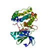
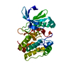
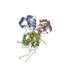
Aurora kinase A / Aurora kinase / Phosphorylase Kinase; domain 1 / Phosphorylase Kinase; domain 1 / Transferase(Phosphotransferase) domain 1 / Transferase(Phosphotransferase); domain 1 / Serine/threonine-protein kinase, active site / Serine/Threonine protein kinases active-site signature. / Protein kinase domain / Serine/Threonine protein kinases, catalytic domain ...Aurora kinase A / Aurora kinase / Phosphorylase Kinase; domain 1 / Phosphorylase Kinase; domain 1 / Transferase(Phosphotransferase) domain 1 / Transferase(Phosphotransferase); domain 1 / Serine/threonine-protein kinase, active site / Serine/Threonine protein kinases active-site signature. / Protein kinase domain / Serine/Threonine protein kinases, catalytic domain / Protein kinase, ATP binding site / Protein kinases ATP-binding region signature. / Immunoglobulins / Protein kinase domain profile. / Protein kinase domain / Protein kinase-like domain superfamily / Immunoglobulin-like / Sandwich / 2-Layer Sandwich / Orthogonal Bundle / Mainly Beta / Mainly Alpha / Alpha Beta 類似検索 - ドメイン・相同性
分子量: 381.432 Da / 分子数: 2 / 由来タイプ: 合成 / 式: C19H23N7O2 / タイプ: SUBJECT OF INVESTIGATION
-
実験情報
-
実験
実験
手法: X線回折 / 使用した結晶の数: 1
-
試料調製
結晶
マシュー密度: 2.28 Å3/Da / 溶媒含有率: 46.05 %
結晶化
温度: 291.15 K / 手法: 蒸気拡散法, シッティングドロップ法 / pH: 5.5 詳細: A 1:1 ratio of protein mixture to mother liquor was obtained by combining 0.5 uL of sample [240 uM deP Aurora A + 1.0 mM AT9283 + 250 uM Mb] with 0.5 uL of mother liquor [0.1 M Bis-Tris, pH 5. ...詳細: A 1:1 ratio of protein mixture to mother liquor was obtained by combining 0.5 uL of sample [240 uM deP Aurora A + 1.0 mM AT9283 + 250 uM Mb] with 0.5 uL of mother liquor [0.1 M Bis-Tris, pH 5.5, 0.2 M magnesium chloride, 19% (w/v) PEG-3350].
-
データ収集
回折
平均測定温度: 100 K
放射光源
由来: シンクロトロン / サイト: ALS / ビームライン: 8.2.1 / 波長: 0.99997 Å
 ムービー
ムービー コントローラー


 データを開く
 基本情報
基本情報 要素
要素 キーワード
キーワード 機能・相同性情報
 Homo sapiens (ヒト)
 X線回折 /
  データ登録者
 米国, 4件
  引用
引用 構造の表示
構造の表示 ダウンロードとリンク
 その他のダウンロード
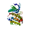


 PDBj
PDBj


 集合体
集合体


 分子量: 381.432 Da / 分子数: 2 / 由来タイプ: 合成 / 式: C19H23N7O2 / タイプ: SUBJECT OF INVESTIGATION
 試料調製
試料調製 解析
解析