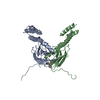
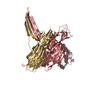
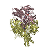
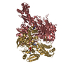
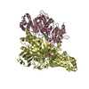
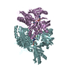
The structure of the I-SspI/DNA complex consists of one protein tetramer bound to a single DNA duplex; the crystallographic asymmetric unit contains one copy of this complex .
-
Components
#1: DNA chain
SyntheticDNA29MER
Mass: 8969.772 Da / Num. of mol.: 1 / Source method: obtained synthetically Details: synthetic construct containing natural homing site of I-SspI6803I
#2: DNA chain
SyntheticDNA29MER
Mass: 8862.685 Da / Num. of mol.: 1 / Source method: obtained synthetically Details: synthetic construct containing natural homing site of I-SspI6803I
#3: Protein
Putativeendonuclease
Mass: 17404.945 Da / Num. of mol.: 4 / Mutation: E11Q,F55K,L16M,L21M Source method: isolated from a genetically manipulated source Source: (gene. exp.) Synechocystis sp. (bacteria) / Strain: PCC 6803 / Plasmid: pET15b / Species (production host): Escherichia coli / Production host: Escherichia coli BL21 (bacteria) / Strain (production host): BL21 / References: UniProt: Q57253
Mass: 18.015 Da / Num. of mol.: 16 / Source method: isolated from a natural source / Formula: H2O
-
Experimental details
-
Experiment
Experiment
Method: X-RAY DIFFRACTION / Number of used crystals: 1
-
Sample preparation
Crystal
Density Matthews: 4.67 Å3/Da / Density % sol: 73.69 %
Crystal grow
Temperature: 277 K / Method: vapor diffusion, hanging drop / pH: 6 Details: 15% to 20% MPD, 100 mM MES buffer, pH 6.0, VAPOR DIFFUSION, HANGING DROP, temperature 277K
Components of the solutions
ID
Name
Crystal-ID
Sol-ID
1
MPD
1
1
2
MES
1
1
3
MPD
1
2
-
Data collection
Diffraction
ID
Mean temperature (K)
Crystal-ID
1
100
1
2
100
1
3
100
1
Diffraction source
Source
Site
Beamline
Type
ID
Wavelength (Å)
Wavelength
SYNCHROTRON
ALS
5.0.2
1
0.979
SYNCHROTRON
ALS
5.0.2
2
0.965
ROTATING ANODE
RIGAKU RU200
3
1.5418
Detector
Type
ID
Detector
Date
ADSC QUANTUM 315
1
CCD
Jun 8, 2006
ADSC QUANTUM 315
2
CCD
Aug 25, 2006
RIGAKU RAXIS V
3
IMAGE PLATE
May 8, 2006
Radiation
ID
Monochromator
Protocol
Monochromatic (M) / Laue (L)
Scattering type
Wavelength-ID
1
Double-crystal, Si(111) liquid N2 cooled
SINGLEWAVELENGTH
M
x-ray
1
2
Double-crystal, Si(111) liquid N2 cooled
SINGLEWAVELENGTH
M
x-ray
1
3
GRAPHITE
SINGLEWAVELENGTH
M
x-ray
1
Radiation wavelength
ID
Wavelength (Å)
Relative weight
1
0.979
1
2
0.965
1
3
1.5418
1
Reflection
D res low: 50 Å
Redundancy (%)
ID
Av σ(I) over netI
Number
Rmerge(I) obs
Χ2
D res high (Å)
Num. obs
% possible obs
7.6
1
10.7
220038
0.083
1.11
3.1
28899
94
9.5
2
6.7
243144
0.152
1.17
3.3
25721
100
Diffraction reflection shell
Highest resolution (Å)
Lowest resolution (Å)
% possible obs (%)
ID
Rmerge(I) obs
Chi squared
Redundancy
8.4
50
96.5
1
0.041
0.953
7.3
6.67
8.4
97.8
1
0.055
1.115
7.3
5.83
6.67
98.7
1
0.08
1.285
7.6
5.3
5.83
98.5
1
0.074
1.259
7.7
4.92
5.3
99.4
1
0.065
1.193
7.7
4.63
4.92
99.3
1
0.067
1.268
7.8
4.4
4.63
99.5
1
0.067
1.33
7.7
4.21
4.4
99.7
1
0.078
1.247
7.8
4.04
4.21
99.7
1
0.09
1.118
7.8
3.91
4.04
99.9
1
0.11
1.059
7.8
3.78
3.91
99.8
1
0.131
1.126
7.8
3.68
3.78
99.9
1
0.143
1.146
7.8
3.58
3.68
99.9
1
0.157
1.075
7.7
3.49
3.58
99.8
1
0.189
1.036
7.7
3.41
3.49
98.4
1
0.192
1.005
7.5
3.34
3.41
94.7
1
0.184
1.006
7.3
3.27
3.34
87.9
1
0.193
1
7.3
3.21
3.27
78.2
1
0.271
0.931
7.3
3.15
3.21
69.3
1
0.298
0.926
7.6
3.1
3.15
61.4
1
0.374
0.907
7.6
8.94
50
99.7
2
0.038
1.165
8.9
7.1
8.94
100
2
0.051
1.031
9.5
6.21
7.1
100
2
0.082
1.112
9
5.64
6.21
100
2
0.107
1.003
9.3
5.24
5.64
100
2
0.107
1.047
9.4
4.93
5.24
100
2
0.109
1.176
9.6
4.68
4.93
100
2
0.126
1.209
9.7
4.48
4.68
100
2
0.125
1.245
9.7
4.31
4.48
100
2
0.14
1.156
9.6
4.16
4.31
99.8
2
0.181
1.124
9.6
4.03
4.16
100
2
0.234
1.128
9.6
3.91
4.03
100
2
0.3
1.187
9.6
3.81
3.91
99.9
2
0.357
1.298
9.5
3.72
3.81
99.9
2
0.388
1.139
9.5
3.63
3.72
100
2
0.419
1.434
9.4
3.55
3.63
100
2
0.559
1.177
9.5
3.48
3.55
99.9
2
0.587
1.175
9.5
3.42
3.48
99.9
2
0.616
1.297
9.4
3.36
3.42
100
2
0.597
1.182
9.4
3.3
3.36
100
2
0.628
1.147
9.4
Reflection
Resolution: 3.1→160.13 Å / Num. obs: 28899 / % possible obs: 94 % / Redundancy: 7.6 % / Rmerge(I) obs: 0.083 / Χ2: 1.111 / Net I/σ(I): 10.7
Reflection shell
Resolution (Å)
Redundancy (%)
Rmerge(I) obs
Num. unique all
Χ2
Diffraction-ID
% possible all
3.1-3.15
7.6
0.374
928
0.907
1,2,3
61.4
3.15-3.21
7.6
0.298
1029
0.926
1,2,3
69.3
3.21-3.27
7.3
0.271
1192
0.931
1,2,3
78.2
3.27-3.34
7.3
0.193
1319
1
1,2,3
87.9
3.34-3.41
7.3
0.184
1420
1.006
1,2,3
94.7
3.41-3.49
7.5
0.192
1491
1.005
1,2,3
98.4
3.49-3.58
7.7
0.189
1531
1.036
1,2,3
99.8
3.58-3.68
7.7
0.157
1511
1.075
1,2,3
99.9
3.68-3.78
7.8
0.143
1529
1.146
1,2,3
99.9
3.78-3.91
7.8
0.131
1498
1.126
1,2,3
99.8
3.91-4.04
7.8
0.11
1523
1.059
1,2,3
99.9
4.04-4.21
7.8
0.09
1549
1.118
1,2,3
99.7
4.21-4.4
7.8
0.078
1515
1.247
1,2,3
99.7
4.4-4.63
7.7
0.067
1533
1.33
1,2,3
99.5
4.63-4.92
7.8
0.067
1516
1.268
1,2,3
99.3
4.92-5.3
7.7
0.065
1529
1.193
1,2,3
99.4
5.3-5.83
7.7
0.074
1543
1.259
1,2,3
98.5
5.83-6.67
7.6
0.08
1543
1.285
1,2,3
98.7
6.67-8.4
7.3
0.055
1566
1.115
1,2,3
97.8
8.4-50
7.3
0.041
1634
0.953
1,2,3
96.5
-
Phasing
Phasing
Method: MAD
Phasing MAD set site
ID
Cartn x (Å)
Cartn y (Å)
Cartn z (Å)
Atom type symbol
B iso
Occupancy
1
-38.362
-15.456
-63.143
SE
71.25
0.74
2
-38.299
-49.252
-50.778
SE
112.21
1.2
3
-33.355
-41.094
-45.778
SE
93.14
1.07
4
-19.776
-34.049
-39.32
SE
140.1
1.28
5
-35.709
-18.056
-67.057
SE
83.37
0.59
6
-28.713
-40.155
-46.549
SE
166.51
1.24
7
-41.981
-24.697
-76.047
SE
90.54
1.12
8
-32.936
-7.045
-58.308
SE
134.88
1.2
Phasing dm
Method: Solvent flattening
-
Processing
Software
Name
Version
Classification
NB
DENZO
datareduction
SCALEPACK
datascaling
SHARP
phasing
DM
6
phasing
REFMAC
refinement
PDB_EXTRACT
2
dataextraction
ADSC
QUANTUM
datacollection
Refinement
Method to determine structure: MAD / Resolution: 3.1→160.13 Å / Cor.coef. Fo:Fc: 0.891 / Cor.coef. Fo:Fc free: 0.845 / SU B: 22.095 / SU ML: 0.402 / Cross valid method: THROUGHOUT / σ(F): 0 / ESU R: 1.024 / ESU R Free: 0.484 / Stereochemistry target values: MAXIMUM LIKELIHOOD
Rfactor
Num. reflection
% reflection
Selection details
Rfree
0.328
1459
5 %
RANDOM
Rwork
0.278
-
-
-
obs
0.281
28898
93.96 %
-
Solvent computation
Ion probe radii: 0.8 Å / Shrinkage radii: 0.8 Å / VDW probe radii: 1.4 Å / Solvent model: MASK
Displacement parameters
Biso mean: 83.07 Å2
Baniso -1
Baniso -2
Baniso -3
1-
3.44 Å2
0 Å2
0 Å2
2-
-
3.44 Å2
0 Å2
3-
-
-
-6.88 Å2
Refinement step
Cycle: LAST / Resolution: 3.1→160.13 Å
Protein
Nucleic acid
Ligand
Solvent
Total
Num. atoms
4540
1189
2
16
5747
Refine LS restraints
Refine-ID
Type
Dev ideal
Dev ideal target
Number
X-RAY DIFFRACTION
r_bond_refined_d
0.007
0.022
5974
X-RAY DIFFRACTION
r_angle_refined_deg
1.889
2.21
8317
X-RAY DIFFRACTION
r_dihedral_angle_1_deg
5.497
5
561
X-RAY DIFFRACTION
r_dihedral_angle_2_deg
36.079
23.244
225
X-RAY DIFFRACTION
r_dihedral_angle_3_deg
19.415
15
803
X-RAY DIFFRACTION
r_dihedral_angle_4_deg
14.392
15
39
X-RAY DIFFRACTION
r_chiral_restr
0.228
0.2
909
X-RAY DIFFRACTION
r_gen_planes_refined
0.002
0.02
4140
X-RAY DIFFRACTION
r_nbd_refined
0.194
0.2
2776
X-RAY DIFFRACTION
r_nbtor_refined
0.302
0.2
3915
X-RAY DIFFRACTION
r_xyhbond_nbd_refined
0.146
0.2
227
X-RAY DIFFRACTION
r_metal_ion_refined
0.111
0.2
6
X-RAY DIFFRACTION
r_symmetry_vdw_refined
0.148
0.2
52
X-RAY DIFFRACTION
r_symmetry_hbond_refined
0.14
0.2
1
X-RAY DIFFRACTION
r_mcbond_it
0.374
1.5
2857
X-RAY DIFFRACTION
r_mcangle_it
0.682
2
4492
X-RAY DIFFRACTION
r_scbond_it
0.609
3
3964
X-RAY DIFFRACTION
r_scangle_it
1.158
4.5
3825
Refine LS restraints NCS
Ens-ID: 1 / Refine-ID: X-RAY DIFFRACTION
Dom-ID
Auth asym-ID
Number
Type
Rms dev position (Å)
Weight position
1
A
500
MEDIUMPOSITIONAL
0.59
0.5
2
B
500
MEDIUMPOSITIONAL
0.42
0.5
3
C
500
MEDIUMPOSITIONAL
0.58
0.5
4
D
500
MEDIUMPOSITIONAL
0.43
0.5
1
A
471
LOOSEPOSITIONAL
1.32
5
2
B
471
LOOSEPOSITIONAL
1.08
5
3
C
471
LOOSEPOSITIONAL
1.23
5
4
D
471
LOOSEPOSITIONAL
1.08
5
1
A
500
MEDIUMTHERMAL
0.55
2
2
B
500
MEDIUMTHERMAL
0.36
2
3
C
500
MEDIUMTHERMAL
0.37
2
4
D
500
MEDIUMTHERMAL
0.45
2
1
A
471
LOOSETHERMAL
1.31
10
2
B
471
LOOSETHERMAL
0.95
10
3
C
471
LOOSETHERMAL
0.97
10
4
D
471
LOOSETHERMAL
1.12
10
LS refinement shell
Resolution: 3.1→3.183 Å / Total num. of bins used: 20
Rfactor
Num. reflection
% reflection
Rfree
0.401
78
-
Rwork
0.328
1305
-
obs
-
1383
62.58 %
+
About Yorodumi
-
News
-
Feb 9, 2022. New format data for meta-information of EMDB entries
New format data for meta-information of EMDB entries
Version 3 of the EMDB header file is now the official format.
The previous official version 1.9 will be removed from the archive.
In the structure databanks used in Yorodumi, some data are registered as the other names, "COVID-19 virus" and "2019-nCoV". Here are the details of the virus and the list of structure data.
Jan 31, 2019. EMDB accession codes are about to change! (news from PDBe EMDB page)
EMDB accession codes are about to change! (news from PDBe EMDB page)
The allocation of 4 digits for EMDB accession codes will soon come to an end. Whilst these codes will remain in use, new EMDB accession codes will include an additional digit and will expand incrementally as the available range of codes is exhausted. The current 4-digit format prefixed with “EMD-” (i.e. EMD-XXXX) will advance to a 5-digit format (i.e. EMD-XXXXX), and so on. It is currently estimated that the 4-digit codes will be depleted around Spring 2019, at which point the 5-digit format will come into force.
The EM Navigator/Yorodumi systems omit the EMD- prefix.
Related info.:Q: What is EMD? / ID/Accession-code notation in Yorodumi/EM Navigator
Yorodumi is a browser for structure data from EMDB, PDB, SASBDB, etc.
This page is also the successor to EM Navigator detail page, and also detail information page/front-end page for Omokage search.
The word "yorodu" (or yorozu) is an old Japanese word meaning "ten thousand". "mi" (miru) is to see.
Related info.:EMDB / PDB / SASBDB / Comparison of 3 databanks / Yorodumi Search / Aug 31, 2016. New EM Navigator & Yorodumi / Yorodumi Papers / Jmol/JSmol / Function and homology information / Changes in new EM Navigator and Yorodumi
 Movie
Movie Controller


 Open data
 Basic information
 Components
 Keywords
 Function and homology information
 X-RAY DIFFRACTION /
  Authors
 Citation
 Structure visualization
 Downloads & links
 Other downloads

 PDBj
PDBj


 Assembly
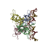

 Mass: 40.078 Da / Num. of mol.: 2 / Source method: obtained synthetically / Formula: Ca
 Mass: 18.015 Da / Num. of mol.: 16 / Source method: isolated from a natural source / Formula: H2O
 Sample preparation
 Processing