 Movie
Movie Controller

[English] 日本語
 Yorodumi
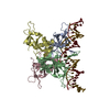
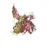
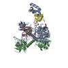
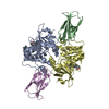
- PDB-4r5g: Crystal structure of the DnaK C-terminus with the inhibitor PET-16 -
+ Open data

- Basic information
| Entry | Database: PDB / ID: 4r5g | ||||||
|---|---|---|---|---|---|---|---|
| Title | Crystal structure of the DnaK C-terminus with the inhibitor PET-16 | ||||||
 Components Components | Chaperone protein DnaK | ||||||
 Keywords Keywords | Chaperone/Chaperone inhibitor / Helical bundle / beta sheets / Chaperone / HSP70/DnaK inhibitors / membrane / Chaperone-Chaperone inhibitor complex | ||||||
| Function / homology |  Function and homology information Function and homology informationstress response to copper ion / sigma factor antagonist activity / protein unfolding / cellular response to unfolded protein / heat shock protein binding / protein folding chaperone / inclusion body / ATP-dependent protein folding chaperone / ADP binding / : ...stress response to copper ion / sigma factor antagonist activity / protein unfolding / cellular response to unfolded protein / heat shock protein binding / protein folding chaperone / inclusion body / ATP-dependent protein folding chaperone / ADP binding / : / response to heat / protein refolding / protein folding / protein-folding chaperone binding / protein-containing complex assembly / DNA replication / ATP hydrolysis activity / protein-containing complex / zinc ion binding / ATP binding / membrane / plasma membrane / cytoplasm / cytosol Similarity search - Function | ||||||
| Biological species |  | ||||||
| Method |  X-RAY DIFFRACTION / SYNCHROTRON / MOLECULAR REPLACEMENT / Resolution: 3.4501 Å X-RAY DIFFRACTION / SYNCHROTRON / MOLECULAR REPLACEMENT / Resolution: 3.4501 Å | ||||||
 Authors Authors | Leu, J.I. / Zhang, P. / Murphy, M.E. / Marmorstein, R. / George, D.L. | ||||||
 Citation Citation | Journal: Acs Chem.Biol. / Year: 2014 Title: Structural Basis for the Inhibition of HSP70 and DnaK Chaperones by Small-Molecule Targeting of a C-Terminal Allosteric Pocket. Authors: Leu, J.I. / Zhang, P. / Murphy, M.E. / Marmorstein, R. / George, D.L. | ||||||
| History |
|
- Structure visualization
| Structure viewer | Molecule: MolmilJmol/JSmol |
|---|
- Downloads & links
-Download
| PDBx/mmCIF format | 4r5g.cif.gz | 87.8 KB | Display | PDBx/mmCIF format |
|---|---|---|---|---|
| PDB format | pdb4r5g.ent.gz | 63.9 KB | Display | PDB format |
| PDBx/mmJSON format | 4r5g.json.gz | Tree view | PDBx/mmJSON format | |
| Others |  Other downloads Other downloads |
-Validation report
| Arichive directory | https://data.pdbj.org/pub/pdb/validation_reports/r5/4r5gftp://data.pdbj.org/pub/pdb/validation_reports/r5/4r5g | HTTPS FTP |
|---|
-Related structure data
| Related structure data | 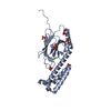 4r5iC 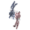 4r5jC 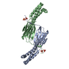 4r5kC 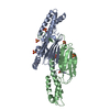 4r5lC 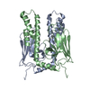 1dkyS S: Starting model for refinement C: citing same article ( |
|---|---|
| Similar structure data |


-Links
 PDBj
PDBj
- Assembly
| Deposited unit | 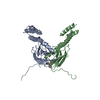
| ||||||||
|---|---|---|---|---|---|---|---|---|---|
| 1 |
| ||||||||
| 2 | 
| ||||||||
| Unit cell |
| ||||||||
| Details | Authors have confirmed dimer by IDT but do not know the proper assembly |
-Components
| #1: Protein | Mass: 25237.375 Da / Num. of mol.: 2 / Fragment: C-terminus of DnaK Source method: isolated from a genetically manipulated source Source: (gene. exp.) #2: Chemical | ChemComp-3JE / |   Mass: 363.411 Da / Num. of mol.: 1 / Fragment: C-terminus of DnaK / Source method: obtained synthetically / Formula: C26H20P Mass: 363.411 Da / Num. of mol.: 1 / Fragment: C-terminus of DnaK / Source method: obtained synthetically / Formula: C26H20P#3: Water | ChemComp-HOH / |  Mass: 18.015 Da / Num. of mol.: 1 / Fragment: HSP70/DnaK inhibitor: PET-16 / Source method: isolated from a natural source / Formula: H2O Mass: 18.015 Da / Num. of mol.: 1 / Fragment: HSP70/DnaK inhibitor: PET-16 / Source method: isolated from a natural source / Formula: H2O |
|---|
-Experimental details
-Experiment
| Experiment | Method: X-RAY DIFFRACTION / Number of used crystals: 1 |
|---|
- Sample preparation
| Crystal | Density Matthews: 2.86 Å3/Da / Density % sol: 56.92 % |
|---|---|
| Crystal grow | Temperature: 293 K / Method: vapor diffusion, hanging drop / pH: 5.5 Details: 1.8M ammonium sulfate, 0.1M Bis-Tris pH 5.5, VAPOR DIFFUSION, HANGING DROP, temperature 293.0K |
-Data collection
| Diffraction | Mean temperature: 100 K |
|---|---|
| Diffraction source | Source: SYNCHROTRON / Site: NSLS  / Beamline: X25 / Wavelength: 1.1 Å / Beamline: X25 / Wavelength: 1.1 Å |
| Detector | Type: ADSC QUANTUM 315 / Detector: CCD / Date: Feb 1, 2013 |
| Radiation | Monochromator: Double silicon(111) crystal monochromator / Protocol: SINGLE WAVELENGTH / Monochromatic (M) / Laue (L): M / Scattering type: x-ray |
| Radiation wavelength | Wavelength: 1.1 Å / Relative weight: 1 |
| Reflection | Resolution: 3.45→45 Å / Num. all: 8306 / Num. obs: 8281 / % possible obs: 99.7 % / Observed criterion σ(F): 1 / Observed criterion σ(I): 1 / Redundancy: 5 % / Rmerge(I) obs: 0.099 / Net I/σ(I): 15 |
| Reflection shell | Resolution: 3.45→3.57 Å / Redundancy: 5.3 % / Rmerge(I) obs: 0.49 / Mean I/σ(I) obs: 2.5 / % possible all: 99.9 |
- Processing
| Software |
| ||||||||||||||||||||||||||||
|---|---|---|---|---|---|---|---|---|---|---|---|---|---|---|---|---|---|---|---|---|---|---|---|---|---|---|---|---|---|
| Refinement | Method to determine structure: MOLECULAR REPLACEMENT Starting model: PDB ENTRY 1DKY Resolution: 3.4501→43.509 Å / SU ML: 0.53 / σ(F): 1.35 / Phase error: 35.79 / Stereochemistry target values: ML
| ||||||||||||||||||||||||||||
| Solvent computation | Shrinkage radii: 0.8 Å / VDW probe radii: 1.1 Å / Solvent model: FLAT BULK SOLVENT MODEL | ||||||||||||||||||||||||||||
| Refinement step | Cycle: LAST / Resolution: 3.4501→43.509 Å
| ||||||||||||||||||||||||||||
| Refine LS restraints |
| ||||||||||||||||||||||||||||
| LS refinement shell |
|
