 Movie
Movie Controller

[English] 日本語
 Yorodumi
- PDB-2w3r: Crystal Structure of Xanthine Dehydrogenase (desulfo form) from R... -
+ Open data

- Basic information
| Entry | Database: PDB / ID: 2w3r | ||||||
|---|---|---|---|---|---|---|---|
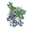
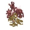
| Title | Crystal Structure of Xanthine Dehydrogenase (desulfo form) from Rhodobacter capsulatus in complex with hypoxanthine | ||||||
 Components Components | (XANTHINE DEHYDROGENASE) x 2 | ||||||
 Keywords Keywords | OXIDOREDUCTASE / XDH / GOUT / IRON / XANTHINE / IRON-SULFUR / MOLYBDENUM COFACTOR / HYPOXANTHINE / METAL-BINDING | ||||||
| Function / homology |  Function and homology information Function and homology information1.1.1.204 / xanthine dehydrogenase activity / molybdenum ion binding / FAD binding / 2 iron, 2 sulfur cluster binding / oxidoreductase activity / iron ion binding Similarity search - Function | ||||||
| Biological species |  RHODOBACTER CAPSULATUS (bacteria) RHODOBACTER CAPSULATUS (bacteria) | ||||||
| Method |  X-RAY DIFFRACTION / SYNCHROTRON / MOLECULAR REPLACEMENT / Resolution: 2.9 Å X-RAY DIFFRACTION / SYNCHROTRON / MOLECULAR REPLACEMENT / Resolution: 2.9 Å | ||||||
 Authors Authors | Dietzel, U. / Kuper, J. / Leimkuhler, S. / Kisker, C. | ||||||
 Citation Citation | Journal: J.Biol.Chem. / Year: 2009 Title: Mechanism of Substrate and Inhibitor Binding of Rhodobacter Capsulatus Xanthine Dehydrogenase. Authors: Dietzel, U. / Kuper, J. / Doebbler, J.A. / Schulte, A. / Truglio, J.J. / Leimkuhler, S. / Kisker, C. | ||||||
| History |
|
- Structure visualization
| Structure viewer | Molecule: MolmilJmol/JSmol |
|---|
- Downloads & links
-Download
| PDBx/mmCIF format | 2w3r.cif.gz | 894.8 KB | Display | PDBx/mmCIF format |
|---|---|---|---|---|
| PDB format | pdb2w3r.ent.gz | 724.2 KB | Display | PDB format |
| PDBx/mmJSON format | 2w3r.json.gz | Tree view | PDBx/mmJSON format | |
| Others |  Other downloads Other downloads |
-Validation report
| Arichive directory | https://data.pdbj.org/pub/pdb/validation_reports/w3/2w3rftp://data.pdbj.org/pub/pdb/validation_reports/w3/2w3r | HTTPS FTP |
|---|
-Related structure data
| Related structure data |  2w3sC 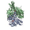 2w54C  2w55C 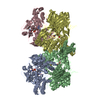 1jroS C: citing same article ( S: Starting model for refinement |
|---|---|
| Similar structure data |
-Links
 PDBj
PDBj


- Assembly
| Deposited unit | 
| ||||||||||||||||||||||||||||||||||||||||||||||||||||||||||||||||||||||||||||||||||||||||||||||||||||||||||||||||||||||||||||||||||||||||||||||||||||
|---|---|---|---|---|---|---|---|---|---|---|---|---|---|---|---|---|---|---|---|---|---|---|---|---|---|---|---|---|---|---|---|---|---|---|---|---|---|---|---|---|---|---|---|---|---|---|---|---|---|---|---|---|---|---|---|---|---|---|---|---|---|---|---|---|---|---|---|---|---|---|---|---|---|---|---|---|---|---|---|---|---|---|---|---|---|---|---|---|---|---|---|---|---|---|---|---|---|---|---|---|---|---|---|---|---|---|---|---|---|---|---|---|---|---|---|---|---|---|---|---|---|---|---|---|---|---|---|---|---|---|---|---|---|---|---|---|---|---|---|---|---|---|---|---|---|---|---|---|---|
| 1 | 
| ||||||||||||||||||||||||||||||||||||||||||||||||||||||||||||||||||||||||||||||||||||||||||||||||||||||||||||||||||||||||||||||||||||||||||||||||||||
| 2 | 
| ||||||||||||||||||||||||||||||||||||||||||||||||||||||||||||||||||||||||||||||||||||||||||||||||||||||||||||||||||||||||||||||||||||||||||||||||||||
| 3 | 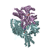
| ||||||||||||||||||||||||||||||||||||||||||||||||||||||||||||||||||||||||||||||||||||||||||||||||||||||||||||||||||||||||||||||||||||||||||||||||||||
| 4 | 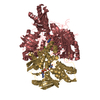
| ||||||||||||||||||||||||||||||||||||||||||||||||||||||||||||||||||||||||||||||||||||||||||||||||||||||||||||||||||||||||||||||||||||||||||||||||||||
| Unit cell |
| ||||||||||||||||||||||||||||||||||||||||||||||||||||||||||||||||||||||||||||||||||||||||||||||||||||||||||||||||||||||||||||||||||||||||||||||||||||
| Noncrystallographic symmetry (NCS) | NCS domain:
NCS domain segments: Component-ID: 1 / Refine code: 4
NCS ensembles :
NCS oper:
|
-Components
-Protein , 2 types, 8 molecules ACEGBDFH
| #1: Protein | Mass: 49420.508 Da / Num. of mol.: 4 Source method: isolated from a genetically manipulated source Source: (gene. exp.) RHODOBACTER CAPSULATUS (bacteria) / Plasmid: PSL207 / Production host: #2: Protein | Mass: 82978.031 Da / Num. of mol.: 4 Source method: isolated from a genetically manipulated source Source: (gene. exp.) RHODOBACTER CAPSULATUS (bacteria) / Plasmid: PSL207 / Production host: |
|---|
-Non-polymers , 7 types, 109 molecules 


| #3: Chemical | ChemComp-FES /  Mass: 175.820 Da / Num. of mol.: 8 / Source method: obtained synthetically / Formula: Fe2S2 Mass: 175.820 Da / Num. of mol.: 8 / Source method: obtained synthetically / Formula: Fe2S2#4: Chemical | ChemComp-FAD /  Mass: 785.550 Da / Num. of mol.: 4 / Source method: obtained synthetically / Formula: C27H33N9O15P2 / Comment: FAD*YM Mass: 785.550 Da / Num. of mol.: 4 / Source method: obtained synthetically / Formula: C27H33N9O15P2 / Comment: FAD*YM#5: Chemical | ChemComp-MTE /  Mass: 395.352 Da / Num. of mol.: 4 / Source method: obtained synthetically / Formula: C10H14N5O6PS2 Mass: 395.352 Da / Num. of mol.: 4 / Source method: obtained synthetically / Formula: C10H14N5O6PS2#6: Chemical | ChemComp-CA /  Mass: 40.078 Da / Num. of mol.: 4 / Source method: obtained synthetically / Formula: Ca Mass: 40.078 Da / Num. of mol.: 4 / Source method: obtained synthetically / Formula: Ca#7: Chemical | ChemComp-HPA /  Mass: 136.111 Da / Num. of mol.: 4 / Source method: obtained synthetically / Formula: C5H4N4O Mass: 136.111 Da / Num. of mol.: 4 / Source method: obtained synthetically / Formula: C5H4N4O#8: Chemical | ChemComp-MOM /  Mass: 144.946 Da / Num. of mol.: 4 / Source method: obtained synthetically / Formula: HMoO3 Mass: 144.946 Da / Num. of mol.: 4 / Source method: obtained synthetically / Formula: HMoO3#9: Water | ChemComp-HOH / | Mass: 18.015 Da / Num. of mol.: 81 / Source method: isolated from a natural source / Formula: H2O |
|---|
-Experimental details
-Experiment
| Experiment | Method: X-RAY DIFFRACTION / Number of used crystals: 1 |
|---|
- Sample preparation
| Crystal | Density Matthews: 3.34 Å3/Da / Density % sol: 59.6 % / Description: NONE |
|---|---|
| Crystal grow | pH: 8.3 Details: 7.5 MM BACL2, 8% PEG 8000, 100 MM TRIS-HCL PH 8.3, 25 MM DTT AND 3% ISOPROPANOL |
-Data collection
| Diffraction | Mean temperature: 100 K |
|---|---|
| Diffraction source | Source: SYNCHROTRON / Site: ESRF  / Beamline: ID23-2 / Wavelength: 0.873 / Beamline: ID23-2 / Wavelength: 0.873 |
| Detector | Type: MARMOSAIC 225 mm CCD / Detector: CCD / Date: Jul 28, 2008 |
| Radiation | Monochromator: SI (111) / Protocol: SINGLE WAVELENGTH / Monochromatic (M) / Laue (L): M / Scattering type: x-ray |
| Radiation wavelength | Wavelength: 0.873 Å / Relative weight: 1 |
| Reflection | Resolution: 2.7→51.03 Å / Num. obs: 183839 / % possible obs: 98.2 % / Observed criterion σ(I): 0 / Redundancy: 1.97 % / Rmerge(I) obs: 0.14 / Net I/σ(I): 6.4 |
| Reflection shell | Resolution: 2.7→2.72 Å / Redundancy: 1.97 % / Rmerge(I) obs: 0.86 / Mean I/σ(I) obs: 0.84 / % possible all: 97.7 |
- Processing
| Software |
| ||||||||||||||||||||||||||||||||||||||||||||||||||||||||||||||||||||||||||||||||||||||||||||||||||||||||||||||||||||||||||||||||||||||||||||||||||||||||||||||||||||||||||||||||||||||
|---|---|---|---|---|---|---|---|---|---|---|---|---|---|---|---|---|---|---|---|---|---|---|---|---|---|---|---|---|---|---|---|---|---|---|---|---|---|---|---|---|---|---|---|---|---|---|---|---|---|---|---|---|---|---|---|---|---|---|---|---|---|---|---|---|---|---|---|---|---|---|---|---|---|---|---|---|---|---|---|---|---|---|---|---|---|---|---|---|---|---|---|---|---|---|---|---|---|---|---|---|---|---|---|---|---|---|---|---|---|---|---|---|---|---|---|---|---|---|---|---|---|---|---|---|---|---|---|---|---|---|---|---|---|---|---|---|---|---|---|---|---|---|---|---|---|---|---|---|---|---|---|---|---|---|---|---|---|---|---|---|---|---|---|---|---|---|---|---|---|---|---|---|---|---|---|---|---|---|---|---|---|---|---|
| Refinement | Method to determine structure: MOLECULAR REPLACEMENT Starting model: PDB ENTRY 1JRO Resolution: 2.9→51.23 Å / Cor.coef. Fo:Fc: 0.895 / Cor.coef. Fo:Fc free: 0.852 / SU B: 19.624 / SU ML: 0.362 / Cross valid method: THROUGHOUT / ESU R Free: 0.426 / Stereochemistry target values: MAXIMUM LIKELIHOOD / Details: HYDROGENS HAVE BEEN ADDED IN THE RIDING POSITIONS.
| ||||||||||||||||||||||||||||||||||||||||||||||||||||||||||||||||||||||||||||||||||||||||||||||||||||||||||||||||||||||||||||||||||||||||||||||||||||||||||||||||||||||||||||||||||||||
| Solvent computation | Ion probe radii: 0.8 Å / Shrinkage radii: 0.8 Å / VDW probe radii: 1.2 Å / Solvent model: MASK | ||||||||||||||||||||||||||||||||||||||||||||||||||||||||||||||||||||||||||||||||||||||||||||||||||||||||||||||||||||||||||||||||||||||||||||||||||||||||||||||||||||||||||||||||||||||
| Displacement parameters | Biso mean: 53.09 Å2
| ||||||||||||||||||||||||||||||||||||||||||||||||||||||||||||||||||||||||||||||||||||||||||||||||||||||||||||||||||||||||||||||||||||||||||||||||||||||||||||||||||||||||||||||||||||||
| Refinement step | Cycle: LAST / Resolution: 2.9→51.23 Å
| ||||||||||||||||||||||||||||||||||||||||||||||||||||||||||||||||||||||||||||||||||||||||||||||||||||||||||||||||||||||||||||||||||||||||||||||||||||||||||||||||||||||||||||||||||||||
| Refine LS restraints |
|