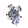
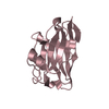
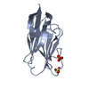
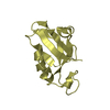
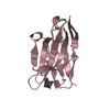
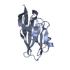
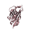
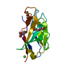
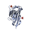
Journal: Science / Year: 2018 Title: Universal protection against influenza infection by a multidomain antibody to influenza hemagglutinin. Authors: Nick S Laursen / Robert H E Friesen / Xueyong Zhu / Mandy Jongeneelen / Sven Blokland / Jan Vermond / Alida van Eijgen / Chan Tang / Harry van Diepen / Galina Obmolova / Marijn van der Neut ...Authors: Nick S Laursen / Robert H E Friesen / Xueyong Zhu / Mandy Jongeneelen / Sven Blokland / Jan Vermond / Alida van Eijgen / Chan Tang / Harry van Diepen / Galina Obmolova / Marijn van der Neut Kolfschoten / David Zuijdgeest / Roel Straetemans / Ryan M B Hoffman / Travis Nieusma / Jesper Pallesen / Hannah L Turner / Steffen M Bernard / Andrew B Ward / Jinquan Luo / Leo L M Poon / Anna P Tretiakova / James M Wilson / Maria P Limberis / Ronald Vogels / Boerries Brandenburg / Joost A Kolkman / Ian A Wilson / Abstract: Broadly neutralizing antibodies against highly variable pathogens have stimulated the design of vaccines and therapeutics. We report the use of diverse camelid single-domain antibodies to influenza ...Broadly neutralizing antibodies against highly variable pathogens have stimulated the design of vaccines and therapeutics. We report the use of diverse camelid single-domain antibodies to influenza virus hemagglutinin to generate multidomain antibodies with impressive breadth and potency. Multidomain antibody MD3606 protects mice against influenza A and B infection when administered intravenously or expressed locally from a recombinant adeno-associated virus vector. Crystal and single-particle electron microscopy structures of these antibodies with hemagglutinins from influenza A and B viruses reveal binding to highly conserved epitopes. Collectively, our findings demonstrate that multidomain antibodies targeting multiple epitopes exhibit enhanced virus cross-reactivity and potency. In combination with adeno-associated virus-mediated gene delivery, they may provide an effective strategy to prevent infection with influenza virus and other highly variable pathogens.
Mass: 12564.941 Da / Num. of mol.: 1 Source method: isolated from a genetically manipulated source Source: (gene. exp.) hybrid / Cell line (production host): HEK293Fexpi / Production host: Homo sapiens (human)
In the structure databanks used in Yorodumi, some data are registered as the other names, "COVID-19 virus" and "2019-nCoV". Here are the details of the virus and the list of structure data.
Jan 31, 2019. EMDB accession codes are about to change! (news from PDBe EMDB page)
EMDB accession codes are about to change! (news from PDBe EMDB page)
The allocation of 4 digits for EMDB accession codes will soon come to an end. Whilst these codes will remain in use, new EMDB accession codes will include an additional digit and will expand incrementally as the available range of codes is exhausted. The current 4-digit format prefixed with “EMD-” (i.e. EMD-XXXX) will advance to a 5-digit format (i.e. EMD-XXXXX), and so on. It is currently estimated that the 4-digit codes will be depleted around Spring 2019, at which point the 5-digit format will come into force.
The EM Navigator/Yorodumi systems omit the EMD- prefix.
Related info.:Q: What is EMD? / ID/Accession-code notation in Yorodumi/EM Navigator
Yorodumi is a browser for structure data from EMDB, PDB, SASBDB, etc.
This page is also the successor to EM Navigator detail page, and also detail information page/front-end page for Omokage search.
The word "yorodu" (or yorozu) is an old Japanese word meaning "ten thousand". "mi" (miru) is to see.
Related info.:EMDB / PDB / SASBDB / Comparison of 3 databanks / Yorodumi Search / Aug 31, 2016. New EM Navigator & Yorodumi / Yorodumi Papers / Jmol/JSmol / Function and homology information / Changes in new EM Navigator and Yorodumi
 Movie
Movie Controller


 Open data
 Basic information
 Components
 Keywords
 Function and homology information
 X-RAY DIFFRACTION /
  Authors
 Citation


 Structure visualization
 Downloads & links
 Other downloads
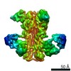


 PDBj
PDBj


 Assembly
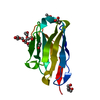
 Homo sapiens (human)
 Mass: 59.044 Da / Num. of mol.: 3 / Source method: obtained synthetically / Formula: C2H3O2
 Mass: 106.120 Da / Num. of mol.: 4 / Source method: obtained synthetically / Formula: C4H10O3
 Mass: 18.015 Da / Num. of mol.: 122 / Source method: isolated from a natural source / Formula: H2O
 Sample preparation
 Processing