 Movie
Movie Controller


+ Open data

- Basic information
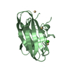
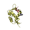
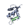
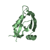
| Entry | Database: PDB / ID: 3dj3 | ||||||
|---|---|---|---|---|---|---|---|
| Title | Crystal Structure of C-terminal Truncated TIP-1 (6-113) | ||||||
 Components Components | Tax1-binding protein 3 | ||||||
 Keywords Keywords | SIGNALING PROTEIN / TIP-1 / PDZ domain / Cytoplasm / Nucleus / Wnt signaling pathway | ||||||
| Function / homology |  Function and homology information Function and homology informationnegative regulation of protein localization to cell surface / regulation of Cdc42 protein signal transduction / negative regulation of Wnt signaling pathway / Rho protein signal transduction / beta-catenin binding / fibrillar center / Wnt signaling pathway / actin cytoskeleton / negative regulation of cell population proliferation / nucleus ...negative regulation of protein localization to cell surface / regulation of Cdc42 protein signal transduction / negative regulation of Wnt signaling pathway / Rho protein signal transduction / beta-catenin binding / fibrillar center / Wnt signaling pathway / actin cytoskeleton / negative regulation of cell population proliferation / nucleus / plasma membrane / cytoplasm Similarity search - Function | ||||||
| Biological species |  | ||||||
| Method |  X-RAY DIFFRACTION / SYNCHROTRON / MOLECULAR REPLACEMENT / Resolution: 2.4 Å X-RAY DIFFRACTION / SYNCHROTRON / MOLECULAR REPLACEMENT / Resolution: 2.4 Å | ||||||
 Authors Authors | Shen, Y. | ||||||
 Citation Citation | Journal: J.Mol.Biol. / Year: 2008 Title: Structural Basis of beta-Catenin Recognition by Tax-interacting Protein-1 Authors: Zhang, J. / Yan, X. / Shi, C. / Yang, X. / Guo, Y. / Tian, C. / Long, J. / Shen, Y. | ||||||
| History |
|
- Structure visualization
| Structure viewer | Molecule: MolmilJmol/JSmol |
|---|
- Downloads & links
-Download
| PDBx/mmCIF format | 3dj3.cif.gz | 89.7 KB | Display | PDBx/mmCIF format |
|---|---|---|---|---|
| PDB format | pdb3dj3.ent.gz | 68.6 KB | Display | PDB format |
| PDBx/mmJSON format | 3dj3.json.gz | Tree view | PDBx/mmJSON format | |
| Others |  Other downloads Other downloads |
-Validation report
| Arichive directory | https://data.pdbj.org/pub/pdb/validation_reports/dj/3dj3ftp://data.pdbj.org/pub/pdb/validation_reports/dj/3dj3 | HTTPS FTP |
|---|
-Related structure data
| Related structure data |  3diwSC  3dj1C S: Starting model for refinement C: citing same article ( |
|---|---|
| Similar structure data |


-Links
 PDBj
PDBj- Assembly
| Deposited unit | 
| ||||||||
|---|---|---|---|---|---|---|---|---|---|
| 1 | 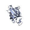
| ||||||||
| 2 | 
| ||||||||
| 3 | 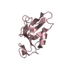
| ||||||||
| 4 | 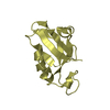
| ||||||||
| Unit cell |
|
-Components
| #1: Protein | Mass: 12508.190 Da / Num. of mol.: 4 / Fragment: c-terminal truncation, UNP residues 1-112 Source method: isolated from a genetically manipulated source Source: (gene. exp.)  #2: Water | ChemComp-HOH / |  Mass: 18.015 Da / Num. of mol.: 103 / Source method: isolated from a natural source / Formula: H2O Mass: 18.015 Da / Num. of mol.: 103 / Source method: isolated from a natural source / Formula: H2O |
|---|
-Experimental details
-Experiment
| Experiment | Method: X-RAY DIFFRACTION / Number of used crystals: 1 |
|---|
- Sample preparation
| Crystal | Density Matthews: 3.08 Å3/Da / Density % sol: 60.12 % |
|---|---|
| Crystal grow | Temperature: 293 K / Method: vapor diffusion, hanging drop / pH: 8 Details: 0.18M DL-malic acid, 25% PEG 3350, pH 8.0, VAPOR DIFFUSION, HANGING DROP, temperature 293K |
-Data collection
| Diffraction | Mean temperature: 100 K |
|---|---|
| Diffraction source | Source: SYNCHROTRON / Site: BSRF  / Beamline: 3W1A / Wavelength: 0.97 Å / Beamline: 3W1A / Wavelength: 0.97 Å |
| Detector | Type: MAR CCD 165 mm / Detector: CCD / Date: Mar 2, 2008 |
| Radiation | Protocol: SINGLE WAVELENGTH / Monochromatic (M) / Laue (L): M / Scattering type: x-ray |
| Radiation wavelength | Wavelength: 0.97 Å / Relative weight: 1 |
| Reflection | Resolution: 2.4→50 Å / Num. all: 23909 / Num. obs: 23790 / % possible obs: 99.5 % / Observed criterion σ(F): 2 / Observed criterion σ(I): 2 / Redundancy: 7.5 % / Biso Wilson estimate: 36.1 Å2 / Rmerge(I) obs: 0.045 / Net I/σ(I): 38 |
| Reflection shell | Resolution: 2.4→2.49 Å / Redundancy: 6.8 % / Rmerge(I) obs: 0.149 / Mean I/σ(I) obs: 10 / Num. unique all: 2374 / % possible all: 98.8 |
- Processing
| Software |
| ||||||||||||||||||||||||||||||||||||
|---|---|---|---|---|---|---|---|---|---|---|---|---|---|---|---|---|---|---|---|---|---|---|---|---|---|---|---|---|---|---|---|---|---|---|---|---|---|
| Refinement | Method to determine structure: MOLECULAR REPLACEMENT Starting model: PDB ENTRY 3DIW Resolution: 2.4→26.03 Å / Rfactor Rfree error: 0.01 / Data cutoff high absF: 602441.81 / Data cutoff low absF: 0 / Isotropic thermal model: RESTRAINED / Cross valid method: THROUGHOUT / σ(F): 0 / Stereochemistry target values: Engh & Huber / Details: BULK SOLVENT MODEL USED
| ||||||||||||||||||||||||||||||||||||
| Solvent computation | Solvent model: FLAT MODEL / Bsol: 34.2045 Å2 / ksol: 0.35 e/Å3 | ||||||||||||||||||||||||||||||||||||
| Displacement parameters | Biso mean: 37.6 Å2
| ||||||||||||||||||||||||||||||||||||
| Refine analyze |
| ||||||||||||||||||||||||||||||||||||
| Refinement step | Cycle: LAST / Resolution: 2.4→26.03 Å
| ||||||||||||||||||||||||||||||||||||
| Refine LS restraints |
| ||||||||||||||||||||||||||||||||||||
| LS refinement shell | Resolution: 2.4→2.55 Å / Rfactor Rfree error: 0.036 / Total num. of bins used: 6
| ||||||||||||||||||||||||||||||||||||
| Xplor file |
|
