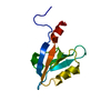
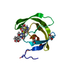
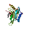
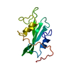
[English] 日本語
Yorodumi
- PDB-3diw: c-terminal beta-catenin bound TIP-1 structure -

+
Open data


ID or keywords:

Loading...

-
Basic information

Entry
Database: PDB / ID: 3diw
Titlec-terminal beta-catenin bound TIP-1 structure
Components
  • Tax1-binding protein 3
  • decameric peptide form Catenin beta-1
KeywordsSIGNALING PROTEIN/CELL ADHESION / beta-catenin / TIP-1 / Tax-interacting protein-1 / PDZ domain / Nucleus / Wnt signaling pathway / SIGNALING PROTEIN-CELL ADHESION COMPLEX
Function / homology
Function and homology information


RHO GTPases Activate Rhotekin and Rhophilins / positive regulation of heparan sulfate proteoglycan biosynthetic process / lung induction / positive regulation of branching involved in lung morphogenesis / cranial ganglion development / renal vesicle formation / renal inner medulla development / renal outer medulla development / nephron tubule formation / beta-catenin-ICAT complex ...RHO GTPases Activate Rhotekin and Rhophilins / positive regulation of heparan sulfate proteoglycan biosynthetic process / lung induction / positive regulation of branching involved in lung morphogenesis / cranial ganglion development / renal vesicle formation / renal inner medulla development / renal outer medulla development / nephron tubule formation / beta-catenin-ICAT complex / CDH11 homotypic and heterotypic interactions / genitalia morphogenesis / embryonic skeletal limb joint morphogenesis / canonical Wnt signaling pathway involved in mesenchymal stem cell differentiation / neural plate development / metanephros morphogenesis / glial cell fate determination / Regulation of CDH19 Expression and Function / regulation of secondary heart field cardioblast proliferation / astrocyte-dopaminergic neuron signaling / oviduct development / beta-catenin-TCF7L2 complex / regulation of nephron tubule epithelial cell differentiation / regulation of timing of anagen / negative regulation of mitotic cell cycle, embryonic / Binding of TCF/LEF:CTNNB1 to target gene promoters / negative regulation of mesenchymal to epithelial transition involved in metanephros morphogenesis / central nervous system vasculogenesis / regulation of centriole-centriole cohesion / RUNX3 regulates WNT signaling / regulation of centromeric sister chromatid cohesion / Regulation of CDH11 function / embryonic axis specification / Specification of the neural plate border / Scrib-APC-beta-catenin complex / lens morphogenesis in camera-type eye / regulation of fibroblast proliferation / beta-catenin-TCF complex / endodermal cell fate commitment / acinar cell differentiation / dorsal root ganglion development / synaptic vesicle clustering / proximal/distal pattern formation / neuron fate determination / Formation of the nephric duct / endothelial tube morphogenesis / negative regulation of protein localization to cell surface / positive regulation of fibroblast growth factor receptor signaling pathway / dorsal/ventral axis specification / sympathetic ganglion development / establishment of blood-retinal barrier / positive regulation of myoblast proliferation / fungiform papilla formation / layer formation in cerebral cortex / positive regulation of endothelial cell differentiation / presynaptic active zone cytoplasmic component / mesenchymal to epithelial transition / hindbrain development / lung epithelial cell differentiation / positive regulation of determination of dorsal identity / positive regulation of skeletal muscle tissue development / fascia adherens / regulation of protein localization to cell surface / ectoderm development / embryonic foregut morphogenesis / hair cell differentiation / detection of muscle stretch / cellular response to indole-3-methanol / smooth muscle cell differentiation / positive regulation of odontoblast differentiation / mesenchymal cell proliferation involved in lung development / histone methyltransferase binding / alpha-catenin binding / regulation of calcium ion import / regulation of epithelial to mesenchymal transition / Germ layer formation at gastrulation / positive regulation of homotypic cell-cell adhesion / establishment of blood-brain barrier / negative regulation of oligodendrocyte differentiation / flotillin complex / apicolateral plasma membrane / epithelial cell differentiation involved in prostate gland development / cranial skeletal system development / positive regulation of epithelial cell proliferation involved in prostate gland development / cell-cell adhesion mediated by cadherin / male genitalia development / Formation of definitive endoderm / regulation of smooth muscle cell proliferation / epithelial cell proliferation involved in prostate gland development / catenin complex / beta-catenin destruction complex / embryonic brain development / oocyte development / lung-associated mesenchyme development / midbrain dopaminergic neuron differentiation / Formation of axial mesoderm / negative regulation of protein sumoylation / Apoptotic cleavage of cell adhesion proteins / LRR FLII-interacting protein 1 (LRRFIP1) activates type I IFN production / Beta-catenin phosphorylation cascade
Similarity search - Function
Tax1-binding protein 3 / Beta-catenin / : / Armadillo/plakoglobin ARM repeat profile. / Armadillo/beta-catenin-like repeat / Armadillo/beta-catenin-like repeats / Armadillo / PDZ domain / Pdz3 Domain / PDZ domain ...Tax1-binding protein 3 / Beta-catenin / : / Armadillo/plakoglobin ARM repeat profile. / Armadillo/beta-catenin-like repeat / Armadillo/beta-catenin-like repeats / Armadillo / PDZ domain / Pdz3 Domain / PDZ domain / PDZ domain profile. / Domain present in PSD-95, Dlg, and ZO-1/2. / PDZ domain / PDZ superfamily / Armadillo-like helical / Armadillo-type fold / Roll / Mainly Beta
Similarity search - Domain/homology
Catenin beta-1 / Tax1-binding protein 3
Similarity search - Component
Biological speciesMus musculus (house mouse)
MethodX-RAY DIFFRACTION / SYNCHROTRON / MOLECULAR REPLACEMENT / Resolution: 2.1 Å
AuthorsShen, Y.
CitationJournal: J.Mol.Biol. / Year: 2008
Title: Structural Basis of beta-Catenin Recognition by Tax-interacting Protein-1
Authors: Zhang, J. / Yan, X. / Shi, C. / Yang, X. / Guo, Y. / Tian, C. / Long, J. / Shen, Y.
History
DepositionJun 21, 2008Deposition site: RCSB / Processing site: PDBJ
Revision 1.0Oct 21, 2008Provider: repository / Type: Initial release
Revision 1.1Jul 13, 2011Group: Version format compliance
Revision 1.2Feb 5, 2014Group: Derived calculations
Revision 1.3Nov 1, 2023Group: Data collection / Database references / Refinement description
Category: chem_comp_atom / chem_comp_bond ...chem_comp_atom / chem_comp_bond / database_2 / pdbx_initial_refinement_model
Item: _database_2.pdbx_DOI / _database_2.pdbx_database_accession

-
Structure visualization

Structure viewerMolecule:
MolmilJmol/JSmol

Downloads & links

-
Assembly

Deposited unit
A: Tax1-binding protein 3
B: Tax1-binding protein 3
C: decameric peptide form Catenin beta-1
D: decameric peptide form Catenin beta-1


Theoretical massNumber of molelcules
Total (without water)29,9244
Polymers29,9244
Non-polymers00
Water3,351186
1
A: Tax1-binding protein 3
C: decameric peptide form Catenin beta-1


Theoretical massNumber of molelcules
Total (without water)14,9622
Polymers14,9622
Non-polymers00
Water362
TypeNameSymmetry operationNumber
identity operation1_555x,y,z1
Buried area1270 Å2
ΔGint-5 kcal/mol
Surface area6820 Å2
MethodPISA
2
B: Tax1-binding protein 3
D: decameric peptide form Catenin beta-1


Theoretical massNumber of molelcules
Total (without water)14,9622
Polymers14,9622
Non-polymers00
Water362
TypeNameSymmetry operationNumber
identity operation1_555x,y,z1
Buried area1110 Å2
ΔGint-6 kcal/mol
Surface area6530 Å2
MethodPISA
Unit cell
Length a, b, c (Å)27.098, 93.571, 107.724
Angle α, β, γ (deg.)90.00, 90.00, 90.00
Int Tables number19
Space group name H-MP212121

-
Components

#1: Protein Tax1-binding protein 3 / Tax interaction protein 1 / TIP-1


Mass: 13739.631 Da / Num. of mol.: 2
Source method: isolated from a genetically manipulated source
Source: (gene. exp.) Mus musculus (house mouse) / Production host: Escherichia coli (E. coli) / References: UniProt: Q9DBG9
#2: Protein/peptide decameric peptide form Catenin beta-1 / Beta-catenin


Mass: 1222.303 Da / Num. of mol.: 2 / Fragment: C-terminal domain, UNP residues 772-781 / Source method: obtained synthetically / Details: This sequence occurs naturally in humans. / References: UniProt: P35222
#3: Water ChemComp-HOH / water


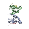
Mass: 18.015 Da / Num. of mol.: 186 / Source method: isolated from a natural source / Formula: H2O

-
Experimental details

-
Experiment

ExperimentMethod: X-RAY DIFFRACTION / Number of used crystals: 1

-
Sample preparation

CrystalDensity Matthews: 2.28 Å3/Da / Density % sol: 46.1 %
Crystal growTemperature: 293 K / Method: vapor diffusion, hanging drop / pH: 4.5
Details: 3.0M sodium formate, 0.1M sodium acetate, pH 4.5, VAPOR DIFFUSION, HANGING DROP, temperature 293K

-
Data collection

DiffractionMean temperature: 100 K
Diffraction sourceSource: SYNCHROTRON / Site: BSRF / Beamline: 3W1A / Wavelength: 0.97 Å
DetectorType: MAR CCD 165 mm / Detector: CCD / Date: Mar 2, 2008
RadiationProtocol: SINGLE WAVELENGTH / Monochromatic (M) / Laue (L): M / Scattering type: x-ray
Radiation wavelengthWavelength: 0.97 Å / Relative weight: 1
ReflectionResolution: 2.1→50 Å / Num. all: 16802 / Num. obs: 16450 / % possible obs: 97.9 % / Observed criterion σ(F): 2 / Observed criterion σ(I): 2 / Redundancy: 6.6 % / Biso Wilson estimate: 15.9 Å2 / Rmerge(I) obs: 0.104 / Net I/σ(I): 13.8
Reflection shellResolution: 2.1→2.18 Å / Redundancy: 4.2 % / Mean I/σ(I) obs: 2.1 / Num. unique all: 1357 / Rsym value: 0.357 / % possible all: 84.4

-
Processing

Software
NameVersionClassification
CNS1.2refinement
HKL-2000data collection
HKL-2000data reduction
SCALEPACKdata scaling
PHASESphasing
RefinementMethod to determine structure: MOLECULAR REPLACEMENT
Starting model: PDB ENTRY 1BE9
Resolution: 2.1→29.96 Å / Rfactor Rfree error: 0.007 / Data cutoff high absF: 167362.42 / Data cutoff low absF: 0 / Isotropic thermal model: RESTRAINED / Cross valid method: THROUGHOUT / σ(F): 0 / Stereochemistry target values: Engh & Huber / Details: BULK SOLVENT MODEL USED
RfactorNum. reflection% reflectionSelection details
Rfree0.265 1569 10 %RANDOM
Rwork0.221 ---
obs0.221 15683 93.4 %-
Solvent computationSolvent model: FLAT MODEL / Bsol: 55.3317 Å2 / ksol: 0.4 e/Å3
Displacement parametersBiso mean: 24.1 Å2
Baniso -1Baniso -2Baniso -3
1--2.57 Å20 Å20 Å2
2--1.48 Å20 Å2
3---1.08 Å2
Refine analyze
FreeObs
Luzzati coordinate error0.32 Å0.25 Å
Luzzati d res low-5 Å
Luzzati sigma a0.25 Å0.21 Å
Refinement stepCycle: LAST / Resolution: 2.1→29.96 Å
ProteinNucleic acidLigandSolventTotal
Num. atoms1816 0 0 186 2002
Refine LS restraints
Refine-IDTypeDev idealDev ideal target
X-RAY DIFFRACTIONc_bond_d0.006
X-RAY DIFFRACTIONc_bond_d_na
X-RAY DIFFRACTIONc_bond_d_prot
X-RAY DIFFRACTIONc_angle_d
X-RAY DIFFRACTIONc_angle_d_na
X-RAY DIFFRACTIONc_angle_d_prot
X-RAY DIFFRACTIONc_angle_deg1.3
X-RAY DIFFRACTIONc_angle_deg_na
X-RAY DIFFRACTIONc_angle_deg_prot
X-RAY DIFFRACTIONc_dihedral_angle_d26.4
X-RAY DIFFRACTIONc_dihedral_angle_d_na
X-RAY DIFFRACTIONc_dihedral_angle_d_prot
X-RAY DIFFRACTIONc_improper_angle_d0.66
X-RAY DIFFRACTIONc_improper_angle_d_na
X-RAY DIFFRACTIONc_improper_angle_d_prot
X-RAY DIFFRACTIONc_mcbond_it1.541.5
X-RAY DIFFRACTIONc_mcangle_it2.52
X-RAY DIFFRACTIONc_scbond_it2.332
X-RAY DIFFRACTIONc_scangle_it3.452.5
LS refinement shellResolution: 2.1→2.23 Å / Rfactor Rfree error: 0.023 / Total num. of bins used: 6
RfactorNum. reflection% reflection
Rfree0.32 196 9.2 %
Rwork0.264 1937 -
obs--78.4 %
Xplor file
Refine-IDSerial noParam fileTopol file
X-RAY DIFFRACTION1protein_rep.paramprotein.top
X-RAY DIFFRACTION2water_rep.paramwater.top

+
About Yorodumi

-
News

-
Feb 9, 2022. New format data for meta-information of EMDB entries

New format data for meta-information of EMDB entries

  • Version 3 of the EMDB header file is now the official format.
  • The previous official version 1.9 will be removed from the archive.

Related info.:EMDB header

External links:wwPDB to switch to version 3 of the EMDB data model

-
Aug 12, 2020. Covid-19 info

Covid-19 info

URL: https://pdbj.org/emnavi/covid19.php

New page: Covid-19 featured information page in EM Navigator.

Related info.:Covid-19 info / Mar 5, 2020. Novel coronavirus structure data

+
Mar 5, 2020. Novel coronavirus structure data

Novel coronavirus structure data

Related info.:Yorodumi Speices / Aug 12, 2020. Covid-19 info

External links:COVID-19 featured content - PDBj / Molecule of the Month (242):Coronavirus Proteases

+
Jan 31, 2019. EMDB accession codes are about to change! (news from PDBe EMDB page)

EMDB accession codes are about to change! (news from PDBe EMDB page)

  • The allocation of 4 digits for EMDB accession codes will soon come to an end. Whilst these codes will remain in use, new EMDB accession codes will include an additional digit and will expand incrementally as the available range of codes is exhausted. The current 4-digit format prefixed with “EMD-” (i.e. EMD-XXXX) will advance to a 5-digit format (i.e. EMD-XXXXX), and so on. It is currently estimated that the 4-digit codes will be depleted around Spring 2019, at which point the 5-digit format will come into force.
  • The EM Navigator/Yorodumi systems omit the EMD- prefix.

Related info.:Q: What is EMD? / ID/Accession-code notation in Yorodumi/EM Navigator

External links:EMDB Accession Codes are Changing Soon! / Contact to PDBj

+
Jul 12, 2017. Major update of PDB

Major update of PDB

  • wwPDB released updated PDB data conforming to the new PDBx/mmCIF dictionary.
  • This is a major update changing the version number from 4 to 5, and with Remediation, in which all the entries are updated.
  • In this update, many items about electron microscopy experimental information are reorganized (e.g. em_software).
  • Now, EM Navigator and Yorodumi are based on the updated data.

External links:wwPDB Remediation / Enriched Model Files Conforming to OneDep Data Standards Now Available in the PDB FTP Archive

-
Yorodumi

Thousand views of thousand structures

  • Yorodumi is a browser for structure data from EMDB, PDB, SASBDB, etc.
  • This page is also the successor to EM Navigator detail page, and also detail information page/front-end page for Omokage search.
  • The word "yorodu" (or yorozu) is an old Japanese word meaning "ten thousand". "mi" (miru) is to see.

Related info.:EMDB / PDB / SASBDB / Comparison of 3 databanks / Yorodumi Search / Aug 31, 2016. New EM Navigator & Yorodumi / Yorodumi Papers / Jmol/JSmol / Function and homology information / Changes in new EM Navigator and Yorodumi

Read more