 Movie
Movie Controller

[English] 日本語
 Yorodumi
- PDB-6cgr: CryoEM structure of herpes simplex virus 1 capsid with associated... -
+ Open data

- Basic information
| Entry | Database: PDB / ID: 6cgr | |||||||||||||||||||||
|---|---|---|---|---|---|---|---|---|---|---|---|---|---|---|---|---|---|---|---|---|---|---|
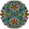
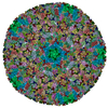
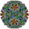
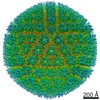
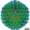
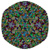
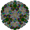
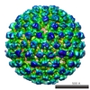
| Title | CryoEM structure of herpes simplex virus 1 capsid with associated tegument protein complexes. | |||||||||||||||||||||
 Components Components |
| |||||||||||||||||||||
 Keywords Keywords | VIRUS / human herpesvirus 1 / Herpes simplex virus type 1 / capsid-associated tegument complex | |||||||||||||||||||||
| Function / homology |  Function and homology information Function and homology informationT=16 icosahedral viral capsid / viral genome packaging / deNEDDylase activity / viral capsid assembly / viral DNA genome replication / viral process / chromosome organization / virion component / viral penetration into host nucleus / viral capsid ...T=16 icosahedral viral capsid / viral genome packaging / deNEDDylase activity / viral capsid assembly / viral DNA genome replication / viral process / chromosome organization / virion component / viral penetration into host nucleus / viral capsid / host cell / symbiont-mediated perturbation of host ubiquitin-like protein modification / cysteine-type deubiquitinase activity / symbiont entry into host cell / host cell nucleus / structural molecule activity / proteolysis / DNA binding Similarity search - Function | |||||||||||||||||||||
| Biological species |   Human herpesvirus 1 (Herpes simplex virus type 1) Human herpesvirus 1 (Herpes simplex virus type 1) | |||||||||||||||||||||
| Method | ELECTRON MICROSCOPY / single particle reconstruction / cryo EM / Resolution: 4.2 Å | |||||||||||||||||||||
 Authors Authors | Dai, X.H. / Zhou, Z.H. | |||||||||||||||||||||
| Funding support |  United States, 6items United States, 6items
| |||||||||||||||||||||
 Citation Citation | Journal: Science / Year: 2018 Title: Structure of the herpes simplex virus 1 capsid with associated tegument protein complexes. Authors: Xinghong Dai / Z Hong Zhou / Abstract: Herpes simplex viruses (HSVs) rely on capsid-associated tegument complex (CATC) for long-range axonal transport of their genome-containing capsids between sites of infection and neuronal cell bodies. ...Herpes simplex viruses (HSVs) rely on capsid-associated tegument complex (CATC) for long-range axonal transport of their genome-containing capsids between sites of infection and neuronal cell bodies. Here we report cryo-electron microscopy structures of the HSV-1 capsid with CATC up to 3.5-angstrom resolution and atomic models of multiple conformers of capsid proteins VP5, VP19c, VP23, and VP26 and tegument proteins pUL17, pUL25, and pUL36. Crowning every capsid vertex are five copies of heteropentameric CATC, each containing a pUL17 monomer supporting the coiled-coil helix bundle of a pUL25 dimer and a pUL36 dimer, thus positioning their flexible domains for potential involvement in nuclear capsid egress and axonal capsid transport. Notwithstanding newly discovered fold conservation between triplex proteins and bacteriophage λ protein gpD and the previously recognized bacteriophage HK97 gp5-like fold in VP5, HSV-1 capsid proteins exhibit extraordinary diversity in forms of domain insertion and conformational polymorphism, not only for interactions with tegument proteins but also for encapsulation of large genomes. | |||||||||||||||||||||
| History |
|
- Structure visualization
| Movie |
Movie viewer |
|---|---|
| Structure viewer | Molecule: MolmilJmol/JSmol |
- Downloads & links
-Download
| PDBx/mmCIF format | 6cgr.cif.gz | 4.7 MB | Display | PDBx/mmCIF format |
|---|---|---|---|---|
| PDB format | pdb6cgr.ent.gz | Display | PDB format | |
| PDBx/mmJSON format | 6cgr.json.gz | Tree view | PDBx/mmJSON format | |
| Others |  Other downloads Other downloads |
-Validation report
| Arichive directory | https://data.pdbj.org/pub/pdb/validation_reports/cg/6cgrftp://data.pdbj.org/pub/pdb/validation_reports/cg/6cgr | HTTPS FTP |
|---|
-Related structure data
| Related structure data | 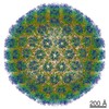 7472MC  7473C M: map data used to model this data C: citing same article ( |
|---|---|
| Similar structure data |


-Links
 PDBj
PDBj
- Assembly
| Deposited unit | 
|
|---|---|
| 1 | x 60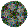
|
| 2 |
|
| 3 | x 5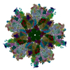
|
| 4 | x 6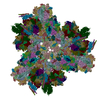
|
| 5 | 
|
| Symmetry | Point symmetry: (Schoenflies symbol: I (icosahedral)) |
-Components
-Protein , 3 types, 33 molecules ABCDEFMNOSTUVWX4GHIJKLPQRYZ012...
| #1: Protein | Mass: 149229.047 Da / Num. of mol.: 16 / Source method: isolated from a natural source Source: (natural) Human herpesvirus 1 (Herpes simplex virus type 1)References: UniProt: G8HBD2 #2: Protein | Mass: 12108.655 Da / Num. of mol.: 15 / Source method: isolated from a natural source Source: (natural) Human herpesvirus 1 (Herpes simplex virus type 1)References: UniProt: Q25BW6 #7: Protein | Mass: 333809.500 Da / Num. of mol.: 2 / Source method: isolated from a natural source Source: (natural) Human herpesvirus 1 (Herpes simplex virus type 1)References: UniProt: G8HBF0, ubiquitinyl hydrolase 1, Hydrolases; Acting on peptide bonds (peptidases); Cysteine endopeptidases |
|---|
-Triplex capsid protein ... , 2 types, 15 molecules 58beh679acdfgij
| #3: Protein | Mass: 50328.281 Da / Num. of mol.: 5 / Source method: isolated from a natural source Source: (natural) Human herpesvirus 1 (Herpes simplex virus type 1)References: UniProt: F8REX2 #4: Protein | Mass: 34301.617 Da / Num. of mol.: 10 / Source method: isolated from a natural source Source: (natural) Human herpesvirus 1 (Herpes simplex virus type 1)References: UniProt: G8H8D9 |
|---|
-Capsid vertex component ... , 2 types, 3 molecules klm
| #5: Protein | Mass: 74699.258 Da / Num. of mol.: 1 / Source method: isolated from a natural source Source: (natural) Human herpesvirus 1 (Herpes simplex virus type 1)References: UniProt: F8RFA1 |
|---|---|
| #6: Protein | Mass: 62736.777 Da / Num. of mol.: 2 / Source method: isolated from a natural source Source: (natural) Human herpesvirus 1 (Herpes simplex virus type 1)References: UniProt: G8HBD8 |
-Details
| Has protein modification | Y |
|---|
-Experimental details
-Experiment
| Experiment | Method: ELECTRON MICROSCOPY |
|---|---|
| EM experiment | Aggregation state: PARTICLE / 3D reconstruction method: single particle reconstruction |
- Sample preparation
| Component | Name: Human herpesvirus 1 strain KOS / Type: VIRUS / Details: Cultured in Vero cells. / Entity ID: all / Source: NATURAL |
|---|---|
| Molecular weight | Value: 200 MDa / Experimental value: NO |
| Source (natural) | Organism: 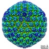 Human herpesvirus 1 strain KOS Human herpesvirus 1 strain KOS |
| Details of virus | Empty: NO / Enveloped: YES / Isolate: STRAIN / Type: VIRION |
| Natural host | Organism: Homo sapiens |
| Virus shell | Name: capsid / Diameter: 1300 nm / Triangulation number (T number): 16 |
| Buffer solution | pH: 7.4 |
| Buffer component | Name: phosphate buffered saline / Formula: PBS |
| Specimen | Embedding applied: NO / Shadowing applied: NO / Staining applied: NO / Vitrification applied: YES |
| Specimen support | Grid material: COPPER / Grid mesh size: 200 divisions/in. / Grid type: Quantifoil R2/1 |
| Vitrification | Instrument: HOMEMADE PLUNGER / Cryogen name: ETHANE / Chamber temperature: 298 K Details: The sample was manually blotted and frozen with a homemade plunger. |
- Electron microscopy imaging
| Experimental equipment |  Model: Titan Krios / Image courtesy: FEI Company |
|---|---|
| Microscopy | Model: FEI TITAN KRIOS |
| Electron gun | Electron source:  FIELD EMISSION GUN / Accelerating voltage: 300 kV / Illumination mode: FLOOD BEAM FIELD EMISSION GUN / Accelerating voltage: 300 kV / Illumination mode: FLOOD BEAM |
| Electron lens | Mode: BRIGHT FIELD / Nominal magnification: 14000 X / Calibrated magnification: 24271 X / Nominal defocus max: 2000 nm / Nominal defocus min: 2000 nm / Cs: 2.7 mm / C2 aperture diameter: 70 µm / Alignment procedure: BASIC |
| Specimen holder | Cryogen: NITROGEN / Specimen holder model: FEI TITAN KRIOS AUTOGRID HOLDER / Temperature (min): 79 K |
| Image recording | Average exposure time: 13 sec. / Electron dose: 25 e/Å2 / Detector mode: SUPER-RESOLUTION / Film or detector model: GATAN K2 SUMMIT (4k x 4k) / Num. of grids imaged: 3 / Num. of real images: 7356 |
| Image scans | Sampling size: 2.5 µm / Width: 7676 / Height: 7420 / Movie frames/image: 26 / Used frames/image: 1-26 |
- Processing
| Software | Name: PHENIX / Version: dev_2875: / Classification: refinement | ||||||||||||||||||||||||
|---|---|---|---|---|---|---|---|---|---|---|---|---|---|---|---|---|---|---|---|---|---|---|---|---|---|
| EM software |
| ||||||||||||||||||||||||
| CTF correction | Type: PHASE FLIPPING AND AMPLITUDE CORRECTION | ||||||||||||||||||||||||
| Particle selection | Num. of particles selected: 45530 Details: Particles were boxed with ETHAN, and then manually examined. | ||||||||||||||||||||||||
| Symmetry | Point symmetry: I (icosahedral) | ||||||||||||||||||||||||
| 3D reconstruction | Resolution: 4.2 Å / Resolution method: FSC 0.143 CUT-OFF / Num. of particles: 28042 / Algorithm: FOURIER SPACE / Symmetry type: POINT | ||||||||||||||||||||||||
| Atomic model building | Protocol: AB INITIO MODEL / Space: REAL | ||||||||||||||||||||||||
| Refine LS restraints |
|
