 Movie
Movie Controller

[English] 日本語
 Yorodumi
- PDB-6bs5: Crystal structure of AMP-PNP-bound bacterial Get3-like A and B in... -
+ Open data

- Basic information
| Entry | Database: PDB / ID: 6bs5 | ||||||
|---|---|---|---|---|---|---|---|
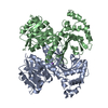
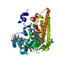
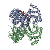
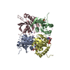
| Title | Crystal structure of AMP-PNP-bound bacterial Get3-like A and B in Mycobacterium tuberculosis | ||||||
 Components Components |
| ||||||
 Keywords Keywords | UNKNOWN FUNCTION / Complex | ||||||
| Function / homology |  Function and homology information Function and homology informationHydrolases; Acting on acid anhydrides; In phosphorus-containing anhydrides / ATP hydrolysis activity / ATP binding / plasma membrane Similarity search - Function | ||||||
| Biological species |   Mycobacterium tuberculosis (bacteria) Mycobacterium tuberculosis (bacteria) | ||||||
| Method |  X-RAY DIFFRACTION / SYNCHROTRON / MOLECULAR REPLACEMENT / Resolution: 3.1 Å X-RAY DIFFRACTION / SYNCHROTRON / MOLECULAR REPLACEMENT / Resolution: 3.1 Å | ||||||
 Authors Authors | Li, H. / Hu, K. / Kovach, A. | ||||||
| Funding support |  United States, 1items United States, 1items
| ||||||
 Citation Citation | Journal: J.Bacteriol. / Year: 2019 Title: Characterization of Guided Entry of Tail-Anchored Proteins 3 Homologues in Mycobacterium tuberculosis. Authors: Hu, K. / Jordan, A.T. / Zhang, S. / Dhabaria, A. / Kovach, A. / Rangel, M.V. / Ueberheide, B. / Li, H. / Darwin, K.H. | ||||||
| History |
|
- Structure visualization
| Structure viewer | Molecule: MolmilJmol/JSmol |
|---|
- Downloads & links
-Download
| PDBx/mmCIF format | 6bs5.cif.gz | 142.9 KB | Display | PDBx/mmCIF format |
|---|---|---|---|---|
| PDB format | pdb6bs5.ent.gz | 108.8 KB | Display | PDB format |
| PDBx/mmJSON format | 6bs5.json.gz | Tree view | PDBx/mmJSON format | |
| Others |  Other downloads Other downloads |
-Validation report
| Arichive directory | https://data.pdbj.org/pub/pdb/validation_reports/bs/6bs5ftp://data.pdbj.org/pub/pdb/validation_reports/bs/6bs5 | HTTPS FTP |
|---|
-Related structure data
| Related structure data |  6bs3SC 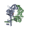 6bs4C S: Starting model for refinement C: citing same article ( |
|---|---|
| Similar structure data |


-Links
 PDBj
PDBj


- Assembly
| Deposited unit | 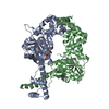
| ||||||||
|---|---|---|---|---|---|---|---|---|---|
| 1 |
| ||||||||
| Unit cell |
|
-Components
| #1: Protein | Mass: 35893.141 Da / Num. of mol.: 1 Source method: isolated from a genetically manipulated source Source: (gene. exp.) Mycobacterium tuberculosis (strain ATCC 25618 / H37Rv) (bacteria)Strain: ATCC 25618 / H37Rv / Gene: Rv3679, MTV025.027 / Production host: |
|---|---|
| #2: Protein | Mass: 42518.656 Da / Num. of mol.: 1 Source method: isolated from a genetically manipulated source Source: (gene. exp.) Mycobacterium tuberculosis (strain ATCC 25618 / H37Rv) (bacteria)Strain: ATCC 25618 / H37Rv / Gene: Rv3680 / Production host: |
| #3: Chemical | ChemComp-ANP /   Mass: 506.196 Da / Num. of mol.: 1 / Source method: obtained synthetically / Formula: C10H17N6O12P3 / Comment: AMP-PNP, energy-carrying molecule analogue*YM Mass: 506.196 Da / Num. of mol.: 1 / Source method: obtained synthetically / Formula: C10H17N6O12P3 / Comment: AMP-PNP, energy-carrying molecule analogue*YM |
| #4: Chemical | ChemComp-MG /   Mass: 24.305 Da / Num. of mol.: 1 / Source method: obtained synthetically / Formula: Mg Mass: 24.305 Da / Num. of mol.: 1 / Source method: obtained synthetically / Formula: Mg |
-Experimental details
-Experiment
| Experiment | Method: X-RAY DIFFRACTION / Number of used crystals: 1 |
|---|
- Sample preparation
| Crystal | Density Matthews: 3.26 Å3/Da / Density % sol: 62.22 % |
|---|---|
| Crystal grow | Temperature: 293 K / Method: vapor diffusion / Details: PEG8000, calcium acetate |
-Data collection
| Diffraction | Mean temperature: 100 K / Serial crystal experiment: N |
|---|---|
| Diffraction source | Source: SYNCHROTRON / Site: APS / Beamline: 31-ID / Wavelength: 1.05969 Å |
| Detector | Type: RAYONIX MX225HE / Detector: CCD / Date: Apr 20, 2017 |
| Radiation | Monochromator: diamond(111) / Protocol: SINGLE WAVELENGTH / Monochromatic (M) / Laue (L): M / Scattering type: x-ray |
| Radiation wavelength | Wavelength: 1.05969 Å / Relative weight: 1 |
| Reflection | Resolution: 3.1→28.282 Å / Num. obs: 18969 / % possible obs: 99.5 % / Redundancy: 6.7 % / Rmerge(I) obs: 0.243 / Net I/σ(I): 8.2 |
| Reflection shell | Resolution: 3.1→3.21 Å / Rmerge(I) obs: 0.958 / Num. unique obs: 2594 |
- Processing
| Software |
| ||||||||||||||||||||||||||||||||||||||||||||||||||||||||
|---|---|---|---|---|---|---|---|---|---|---|---|---|---|---|---|---|---|---|---|---|---|---|---|---|---|---|---|---|---|---|---|---|---|---|---|---|---|---|---|---|---|---|---|---|---|---|---|---|---|---|---|---|---|---|---|---|---|
| Refinement | Method to determine structure: MOLECULAR REPLACEMENT Starting model: PDB entry 6BS3 Resolution: 3.1→28.282 Å / SU ML: 0.32 / Cross valid method: FREE R-VALUE / σ(F): 1.36 / Phase error: 22.46
| ||||||||||||||||||||||||||||||||||||||||||||||||||||||||
| Solvent computation | Shrinkage radii: 0.9 Å / VDW probe radii: 1.11 Å | ||||||||||||||||||||||||||||||||||||||||||||||||||||||||
| Refinement step | Cycle: LAST / Resolution: 3.1→28.282 Å
| ||||||||||||||||||||||||||||||||||||||||||||||||||||||||
| Refine LS restraints |
| ||||||||||||||||||||||||||||||||||||||||||||||||||||||||
| LS refinement shell |
|
