 Movie
Movie Controller


+ Open data

- Basic information
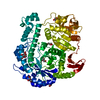
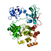
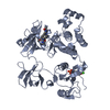
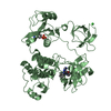
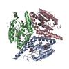
| Entry | Database: PDB / ID: 6bk6 | ||||||
|---|---|---|---|---|---|---|---|
| Title | Crystal structure of Hendra virus matrix protein | ||||||
 Components Components | Hendra virus matrix protein | ||||||
 Keywords Keywords | VIRAL PROTEIN / Hendra virus / viral assembly | ||||||
| Function / homology |  Function and homology information Function and homology informationvirion assembly / virion component / structural constituent of virion / host cell cytoplasm / host cell nucleus Similarity search - Function | ||||||
| Biological species |  Hendra henipavirus Hendra henipavirus | ||||||
| Method |  X-RAY DIFFRACTION / SYNCHROTRON / MOLECULAR REPLACEMENT / Resolution: 2.5 Å X-RAY DIFFRACTION / SYNCHROTRON / MOLECULAR REPLACEMENT / Resolution: 2.5 Å | ||||||
 Authors Authors | Liu, Y.C. | ||||||
 Citation Citation | Journal: J. Virol. / Year: 2018 Title: Electrostatic Interactions between Hendra Virus Matrix Proteins Are Required for Efficient Virus-Like-Particle Assembly. Authors: Liu, Y.C. / Grusovin, J. / Adams, T.E. | ||||||
| History |
|
- Structure visualization
| Structure viewer | Molecule: MolmilJmol/JSmol |
|---|
- Downloads & links
-Download
| PDBx/mmCIF format | 6bk6.cif.gz | 75.4 KB | Display | PDBx/mmCIF format |
|---|---|---|---|---|
| PDB format | pdb6bk6.ent.gz | 53.5 KB | Display | PDB format |
| PDBx/mmJSON format | 6bk6.json.gz | Tree view | PDBx/mmJSON format | |
| Others |  Other downloads Other downloads |
-Validation report
| Arichive directory | https://data.pdbj.org/pub/pdb/validation_reports/bk/6bk6ftp://data.pdbj.org/pub/pdb/validation_reports/bk/6bk6 | HTTPS FTP |
|---|
-Related structure data
| Related structure data | 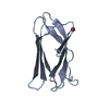 4gioS S: Starting model for refinement |
|---|---|
| Similar structure data |


-Links
 PDBj
PDBj
- Assembly
| Deposited unit | 
| ||||||||
|---|---|---|---|---|---|---|---|---|---|
| 1 | 
| ||||||||
| Unit cell |
|
-Components
| #1: Protein | Mass: 42243.707 Da / Num. of mol.: 1 Source method: isolated from a genetically manipulated source Source: (gene. exp.) Hendra henipavirus / Production host:   Spodoptera frugiperda (fall armyworm) / References: UniProt: F4YH69, UniProt: O89341*PLUS Spodoptera frugiperda (fall armyworm) / References: UniProt: F4YH69, UniProt: O89341*PLUS |
|---|---|
| #2: Chemical | ChemComp-ACT /   Mass: 59.044 Da / Num. of mol.: 1 / Source method: obtained synthetically / Formula: C2H3O2 Mass: 59.044 Da / Num. of mol.: 1 / Source method: obtained synthetically / Formula: C2H3O2 |
| #3: Water | ChemComp-HOH /  Mass: 18.015 Da / Num. of mol.: 62 / Source method: isolated from a natural source / Formula: H2O Mass: 18.015 Da / Num. of mol.: 62 / Source method: isolated from a natural source / Formula: H2O |
| Has protein modification | Y |
-Experimental details
-Experiment
| Experiment | Method: X-RAY DIFFRACTION / Number of used crystals: 1 |
|---|
- Sample preparation
| Crystal | Density Matthews: 2.27 Å3/Da / Density % sol: 45.88 % |
|---|---|
| Crystal grow | Temperature: 293 K / Method: vapor diffusion, hanging drop Details: 0.2 M Ca-Acetate, 0.1 M imadazole pH 7.5, 5% PEG 8000 |
-Data collection
| Diffraction | Mean temperature: 200 K |
|---|---|
| Diffraction source | Source: SYNCHROTRON / Site: Australian Synchrotron  / Beamline: MX2 / Wavelength: 0.953735 Å / Beamline: MX2 / Wavelength: 0.953735 Å |
| Detector | Type: DECTRIS EIGER X 16M / Detector: PIXEL / Date: Mar 30, 2017 |
| Radiation | Protocol: SINGLE WAVELENGTH / Monochromatic (M) / Laue (L): M / Scattering type: x-ray |
| Radiation wavelength | Wavelength: 0.953735 Å / Relative weight: 1 |
| Reflection | Resolution: 2.5→52.291 Å / Num. obs: 14447 / % possible obs: 98.8 % / Redundancy: 5.7 % / Net I/σ(I): 11.2 |
| Reflection shell | Resolution: 2.5→2.6 Å / Rmerge(I) obs: 0.425 / Mean I/σ(I) obs: 3.9 / Num. unique all: 1583 / % possible all: 99.7 |
- Processing
| Software |
| ||||||||||||||||||||||||||||||||||||||||||
|---|---|---|---|---|---|---|---|---|---|---|---|---|---|---|---|---|---|---|---|---|---|---|---|---|---|---|---|---|---|---|---|---|---|---|---|---|---|---|---|---|---|---|---|
| Refinement | Method to determine structure: MOLECULAR REPLACEMENT Starting model: 4GIO Resolution: 2.5→52.291 Å / SU ML: 0.26 / Cross valid method: FREE R-VALUE / σ(F): 1.34 / Phase error: 25.51
| ||||||||||||||||||||||||||||||||||||||||||
| Solvent computation | Shrinkage radii: 0.9 Å / VDW probe radii: 1.11 Å | ||||||||||||||||||||||||||||||||||||||||||
| Refinement step | Cycle: LAST / Resolution: 2.5→52.291 Å
| ||||||||||||||||||||||||||||||||||||||||||
| Refine LS restraints |
| ||||||||||||||||||||||||||||||||||||||||||
| LS refinement shell |
|
