 Movie
Movie Controller

[English] 日本語
 Yorodumi
- PDB-6y2k: Crystal structure of beta-galactosidase from the psychrophilic Ma... -
+ Open data

- Basic information
| Entry | Database: PDB / ID: 6y2k | ||||||
|---|---|---|---|---|---|---|---|
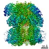
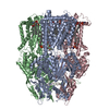
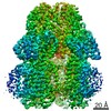
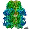
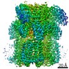
| Title | Crystal structure of beta-galactosidase from the psychrophilic Marinomonas ef1 | ||||||
 Components Components | beta-galactosidase | ||||||
 Keywords Keywords | HYDROLASE / glycosyl hydrolase / psychrophilic enzyme / hexameric structure / cold adaptation / enzyme kinetics / cooperativity | ||||||
| Function / homology |  Function and homology information Function and homology informationClass I glutamine amidotransferase (GATase) domain / Golgi alpha-mannosidase II / Glycosidases / TIM Barrel / Alpha-Beta Barrel / Immunoglobulin-like / Sandwich / Rossmann fold / 3-Layer(aba) Sandwich / Mainly Beta / Alpha Beta Similarity search - Domain/homology | ||||||
| Biological species |  Marinomonas sp. ef1 (bacteria) Marinomonas sp. ef1 (bacteria) | ||||||
| Method |  X-RAY DIFFRACTION / SYNCHROTRON / MOLECULAR REPLACEMENT / Resolution: 1.9 Å X-RAY DIFFRACTION / SYNCHROTRON / MOLECULAR REPLACEMENT / Resolution: 1.9 Å | ||||||
 Authors Authors | Mangiagalli, M. / Lapi, M. / Maione, S. / Orlando, M. / Brocca, S. / Pesce, A. / Barbiroli, A. / Pucciarelli, S. / Camilloni, C. / Lotti, M. | ||||||
 Citation Citation | Journal: Febs J. / Year: 2021 Title: The co-existence of cold activity and thermal stability in an Antarctic GH42 beta-galactosidase relies on its hexameric quaternary arrangement. Authors: Mangiagalli, M. / Lapi, M. / Maione, S. / Orlando, M. / Brocca, S. / Pesce, A. / Barbiroli, A. / Camilloni, C. / Pucciarelli, S. / Lotti, M. / Nardini, M. | ||||||
| History |
|
- Structure visualization
| Structure viewer | Molecule: MolmilJmol/JSmol |
|---|
- Downloads & links
-Download
| PDBx/mmCIF format | 6y2k.cif.gz | 171.2 KB | Display | PDBx/mmCIF format |
|---|---|---|---|---|
| PDB format | pdb6y2k.ent.gz | 132.6 KB | Display | PDB format |
| PDBx/mmJSON format | 6y2k.json.gz | Tree view | PDBx/mmJSON format | |
| Others |  Other downloads Other downloads |
-Validation report
| Arichive directory | https://data.pdbj.org/pub/pdb/validation_reports/y2/6y2kftp://data.pdbj.org/pub/pdb/validation_reports/y2/6y2k | HTTPS FTP |
|---|
-Related structure data
| Related structure data |  1kwgS S: Starting model for refinement |
|---|---|
| Similar structure data |


-Links
 PDBj
PDBj- Assembly
| Deposited unit | 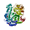
| ||||||||||||
|---|---|---|---|---|---|---|---|---|---|---|---|---|---|
| 1 | x 6
| ||||||||||||
| Unit cell |
| ||||||||||||
| Components on special symmetry positions |
|
-Components
| #1: Protein | Mass: 75248.938 Da / Num. of mol.: 1 Source method: isolated from a genetically manipulated source Source: (gene. exp.) Marinomonas sp. ef1 (bacteria) / Production host: | ||||||
|---|---|---|---|---|---|---|---|
| #2: Chemical |   Mass: 35.453 Da / Num. of mol.: 2 / Source method: obtained synthetically / Formula: Cl / Feature type: SUBJECT OF INVESTIGATION Mass: 35.453 Da / Num. of mol.: 2 / Source method: obtained synthetically / Formula: Cl / Feature type: SUBJECT OF INVESTIGATION#3: Chemical | ChemComp-GOL /   Mass: 92.094 Da / Num. of mol.: 10 / Source method: obtained synthetically / Formula: C3H8O3 Mass: 92.094 Da / Num. of mol.: 10 / Source method: obtained synthetically / Formula: C3H8O3#4: Water | ChemComp-HOH / |  Mass: 18.015 Da / Num. of mol.: 978 / Source method: isolated from a natural source / Formula: H2O Mass: 18.015 Da / Num. of mol.: 978 / Source method: isolated from a natural source / Formula: H2OHas ligand of interest | Y | |
-Experimental details
-Experiment
| Experiment | Method: X-RAY DIFFRACTION / Number of used crystals: 1 |
|---|
- Sample preparation
| Crystal | Density Matthews: 4 Å3/Da / Density % sol: 69.6 % |
|---|---|
| Crystal grow | Temperature: 294 K / Method: vapor diffusion, sitting drop / pH: 5.6 Details: 0.5 M NaCl, 100 mM Na-citrate, 2% ethylene imine polymer. |
-Data collection
| Diffraction | Mean temperature: 100 K / Serial crystal experiment: N |
|---|---|
| Diffraction source | Source: SYNCHROTRON / Site: ESRF  / Beamline: ID14-4 / Wavelength: 0.87313 Å / Beamline: ID14-4 / Wavelength: 0.87313 Å |
| Detector | Type: DECTRIS PILATUS3 S 6M / Detector: PIXEL / Date: Jun 14, 2018 |
| Radiation | Protocol: SINGLE WAVELENGTH / Monochromatic (M) / Laue (L): M / Scattering type: x-ray |
| Radiation wavelength | Wavelength: 0.87313 Å / Relative weight: 1 |
| Reflection | Resolution: 1.9→116.29 Å / Num. obs: 96335 / % possible obs: 100 % / Redundancy: 29.4 % / Rmerge(I) obs: 0.255 / Net I/σ(I): 10.9 |
| Reflection shell | Resolution: 1.9→2 Å / Redundancy: 33.6 % / Rmerge(I) obs: 0.674 / Num. unique obs: 3405 / % possible all: 99.7 |
- Processing
| Software |
| ||||||||||||||||||||||||||||||||||||||||||||||||||||||||||||
|---|---|---|---|---|---|---|---|---|---|---|---|---|---|---|---|---|---|---|---|---|---|---|---|---|---|---|---|---|---|---|---|---|---|---|---|---|---|---|---|---|---|---|---|---|---|---|---|---|---|---|---|---|---|---|---|---|---|---|---|---|---|
| Refinement | Method to determine structure: MOLECULAR REPLACEMENT Starting model: 1KWG Resolution: 1.9→116.29 Å / Cor.coef. Fo:Fc: 0.962 / Cor.coef. Fo:Fc free: 0.948 / SU B: 2.462 / SU ML: 0.068 / Cross valid method: THROUGHOUT / σ(F): 0 / ESU R: 0.103 / ESU R Free: 0.1
| ||||||||||||||||||||||||||||||||||||||||||||||||||||||||||||
| Solvent computation | Ion probe radii: 0.8 Å / Shrinkage radii: 0.8 Å / VDW probe radii: 1.2 Å | ||||||||||||||||||||||||||||||||||||||||||||||||||||||||||||
| Displacement parameters | Biso max: 133.35 Å2 / Biso mean: 20.48 Å2 / Biso min: 7.36 Å2
| ||||||||||||||||||||||||||||||||||||||||||||||||||||||||||||
| Refinement step | Cycle: final / Resolution: 1.9→116.29 Å
| ||||||||||||||||||||||||||||||||||||||||||||||||||||||||||||
| Refine LS restraints |
| ||||||||||||||||||||||||||||||||||||||||||||||||||||||||||||
| LS refinement shell | Resolution: 1.9→1.949 Å / Rfactor Rfree error: 0 / Total num. of bins used: 20
|
