- PDB-2rdd: X-ray crystal structure of AcrB in complex with a novel transmemb... -
+
Open data
ID or keywords:
Loading...
-
Basic information
Entry
Database: PDB / ID: 2rdd
Title
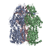
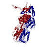
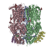
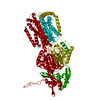
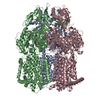
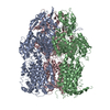
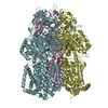
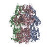
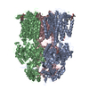
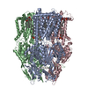
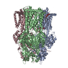
X-ray crystal structure of AcrB in complex with a novel transmembrane helix.
Components
Acriflavine resistance protein B
UPF0092 membrane protein yajC
Keywords
Membrane protein/TRANSPORT PROTEIN / DRUG RESISTANCE / MULTIDRUG EFFLUX / TRANSPORTER / ANTIPORTER / MEMBRANE PROTEIN / NOVEL TRANSMEMBRANE HELIX / ACRB / YAJC / Inner membrane / Membrane protein-TRANSPORT PROTEIN COMPLEX
Function / homology
Function and homology information
protein transport by the Sec complex / alkane transmembrane transporter activity / alkane transport / enterobactin transport / enterobactin transmembrane transporter activity / xenobiotic detoxification by transmembrane export across the cell outer membrane / periplasmic side of plasma membrane / efflux pump complex / Iron assimilation using enterobactin / bile acid transmembrane transporter activity ...protein transport by the Sec complex / alkane transmembrane transporter activity / alkane transport / enterobactin transport / enterobactin transmembrane transporter activity / xenobiotic detoxification by transmembrane export across the cell outer membrane / periplasmic side of plasma membrane / efflux pump complex / Iron assimilation using enterobactin / bile acid transmembrane transporter activity / Antimicrobial resistance / bile acid and bile salt transport / Secretion of toxins / xenobiotic transmembrane transporter activity / efflux transmembrane transporter activity / fatty acid transport / xenobiotic transport / response to toxic substance / response to xenobiotic stimulus / response to antibiotic / membrane / identical protein binding / plasma membrane Similarity search - Function
Resolution: 3.5→50 Å / Cross valid method: THROUGHOUT / Stereochemistry target values: Engh & Huber Details: Polyalanine model of Chain B (YajC) except for five residues that showed positive side chain electron density(above 3.0 sigma level) in the Fobs-Fcalc density map calculated with an all-polyalanine model.
Rfactor
Num. reflection
% reflection
Selection details
Rfree
0.317
1181
-
Random
Rwork
0.279
-
-
-
obs
-
23525
88 %
-
Displacement parameters
Biso mean: 65.6 Å2
Refine analyze
Free
Obs
Luzzati coordinate error
0.65 Å
0.56 Å
Luzzati d res low
-
5 Å
Luzzati sigma a
1.04 Å
0.77 Å
Refinement step
Cycle: LAST / Resolution: 3.5→50 Å
Protein
Nucleic acid
Ligand
Solvent
Total
Num. atoms
7980
0
48
0
8028
Refine LS restraints
Refine-ID
Type
Dev ideal
X-RAY DIFFRACTION
c_bond_d
0.011
X-RAY DIFFRACTION
c_angle_deg
1.9
X-RAY DIFFRACTION
c_dihedral_angle_d
22.7
X-RAY DIFFRACTION
c_improper_angle_d
1.3
+
About Yorodumi
-
News
-
Feb 9, 2022. New format data for meta-information of EMDB entries
New format data for meta-information of EMDB entries
Version 3 of the EMDB header file is now the official format.
The previous official version 1.9 will be removed from the archive.
In the structure databanks used in Yorodumi, some data are registered as the other names, "COVID-19 virus" and "2019-nCoV". Here are the details of the virus and the list of structure data.
Jan 31, 2019. EMDB accession codes are about to change! (news from PDBe EMDB page)
EMDB accession codes are about to change! (news from PDBe EMDB page)
The allocation of 4 digits for EMDB accession codes will soon come to an end. Whilst these codes will remain in use, new EMDB accession codes will include an additional digit and will expand incrementally as the available range of codes is exhausted. The current 4-digit format prefixed with “EMD-” (i.e. EMD-XXXX) will advance to a 5-digit format (i.e. EMD-XXXXX), and so on. It is currently estimated that the 4-digit codes will be depleted around Spring 2019, at which point the 5-digit format will come into force.
The EM Navigator/Yorodumi systems omit the EMD- prefix.
Related info.:Q: What is EMD? / ID/Accession-code notation in Yorodumi/EM Navigator
Yorodumi is a browser for structure data from EMDB, PDB, SASBDB, etc.
This page is also the successor to EM Navigator detail page, and also detail information page/front-end page for Omokage search.
The word "yorodu" (or yorozu) is an old Japanese word meaning "ten thousand". "mi" (miru) is to see.
Related info.:EMDB / PDB / SASBDB / Comparison of 3 databanks / Yorodumi Search / Aug 31, 2016. New EM Navigator & Yorodumi / Yorodumi Papers / Jmol/JSmol / Function and homology information / Changes in new EM Navigator and Yorodumi
 Movie
Movie Controller

 Yorodumi
 Open data
 Basic information
 Components
 Keywords
 Function and homology information
 X-RAY DIFFRACTION /
  Authors
 Citation
 Structure visualization
 Downloads & links
 Other downloads
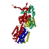


 PDBj
PDBj
 Assembly


 Mass: 349.405 Da / Num. of mol.: 2 / Source method: obtained synthetically / Formula: C16H19N3O4S / Comment: antibiotic*YM
 Sample preparation
 / Beamline: ID14-2 / Wavelength: 0.933 Å
 Processing