 Movie
Movie Controller

[English] 日本語
 Yorodumi
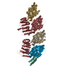
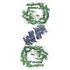
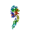
- PDB-6bbn: Crystal structure of a curved tubulin complex induced by the kine... -
+ Open data

- Basic information
| Entry | Database: PDB / ID: 6bbn | |||||||||
|---|---|---|---|---|---|---|---|---|---|---|
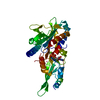
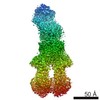
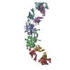
| Title | Crystal structure of a curved tubulin complex induced by the kinesin-13 Kif2A | |||||||||
 Components Components |
| |||||||||
 Keywords Keywords | MOTOR PROTEIN / Kinesin / Kif2A / microtubules / tubulin / kinesin-13 | |||||||||
| Function / homology |  Function and homology information Function and homology informationsperm annulus / centriolar subdistal appendage / Kinesins / positive regulation of axon guidance / microtubule depolymerization / microtubule motor activity / kinesin complex / COPI-dependent Golgi-to-ER retrograde traffic / microtubule-based movement / cytoskeletal motor activity ...sperm annulus / centriolar subdistal appendage / Kinesins / positive regulation of axon guidance / microtubule depolymerization / microtubule motor activity / kinesin complex / COPI-dependent Golgi-to-ER retrograde traffic / microtubule-based movement / cytoskeletal motor activity / mitotic spindle assembly / microtubule-based process / cytoplasmic microtubule / cellular response to interleukin-4 / Amplification of signal from unattached kinetochores via a MAD2 inhibitory signal / MHC class II antigen presentation / centriole / Mitotic Prometaphase / regulation of cell migration / EML4 and NUDC in mitotic spindle formation / Resolution of Sister Chromatid Cohesion / mitotic spindle organization / sperm principal piece / RHO GTPases Activate Formins / microtubule cytoskeleton organization / structural constituent of cytoskeleton / neuron migration / spindle / spindle pole / mitotic spindle / Separation of Sister Chromatids / nervous system development / mitotic cell cycle / double-stranded RNA binding / sperm midpiece / microtubule cytoskeleton / microtubule binding / microtubule / Hydrolases; Acting on acid anhydrides; Acting on GTP to facilitate cellular and subcellular movement / cell differentiation / cilium / ciliary basal body / nuclear body / protein heterodimerization activity / cell division / GTPase activity / centrosome / ubiquitin protein ligase binding / nucleolus / GTP binding / ATP hydrolysis activity / nucleoplasm / ATP binding / membrane / metal ion binding / cytosol / cytoplasm Similarity search - Function | |||||||||
| Biological species |   Homo sapiens (human) Homo sapiens (human) | |||||||||
| Method |  X-RAY DIFFRACTION / SYNCHROTRON / MOLECULAR REPLACEMENT / Resolution: 3.514 Å X-RAY DIFFRACTION / SYNCHROTRON / MOLECULAR REPLACEMENT / Resolution: 3.514 Å | |||||||||
 Authors Authors | Allingham, J.S. / Trofimova, D. | |||||||||
| Funding support |  Canada, 2items Canada, 2items
| |||||||||
 Citation Citation | Journal: Nat Commun / Year: 2018 Title: Ternary complex of Kif2A-bound tandem tubulin heterodimers represents a kinesin-13-mediated microtubule depolymerization reaction intermediate. Authors: Trofimova, D. / Paydar, M. / Zara, A. / Talje, L. / Kwok, B.H. / Allingham, J.S. | |||||||||
| History |
|
- Structure visualization
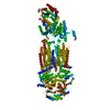
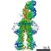
| Structure viewer | Molecule: MolmilJmol/JSmol |
|---|
- Downloads & links
-Download
| PDBx/mmCIF format | 6bbn.cif.gz | 460.3 KB | Display | PDBx/mmCIF format |
|---|---|---|---|---|
| PDB format | pdb6bbn.ent.gz | 364.9 KB | Display | PDB format |
| PDBx/mmJSON format | 6bbn.json.gz | Tree view | PDBx/mmJSON format | |
| Others |  Other downloads Other downloads |
-Validation report
| Arichive directory | https://data.pdbj.org/pub/pdb/validation_reports/bb/6bbnftp://data.pdbj.org/pub/pdb/validation_reports/bb/6bbn | HTTPS FTP |
|---|
-Related structure data
-Links
 PDBj
PDBj


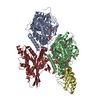
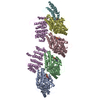
- Assembly
| Deposited unit | 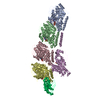
| ||||||||
|---|---|---|---|---|---|---|---|---|---|
| 1 |
| ||||||||
| Unit cell |
|
-Components
-Protein , 4 types, 6 molecules ACBDPE
| #1: Protein | Mass: 50204.445 Da / Num. of mol.: 2 / Source method: isolated from a natural source / Source: (natural) #2: Protein | Mass: 49999.887 Da / Num. of mol.: 2 / Source method: isolated from a natural source / Source: (natural) #3: Protein | | Mass: 17911.244 Da / Num. of mol.: 1 Source method: isolated from a genetically manipulated source Source: (gene. exp.) #4: Protein | | Mass: 47918.902 Da / Num. of mol.: 1 Source method: isolated from a genetically manipulated source Source: (gene. exp.) Homo sapiens (human) / Gene: KIF2A, KIF2, KNS2 / Production host: |
|---|
-Non-polymers , 5 types, 10 molecules 


| #5: Chemical |  Mass: 523.180 Da / Num. of mol.: 2 / Source method: obtained synthetically / Formula: C10H16N5O14P3 / Comment: GTP, energy-carrying molecule*YM Mass: 523.180 Da / Num. of mol.: 2 / Source method: obtained synthetically / Formula: C10H16N5O14P3 / Comment: GTP, energy-carrying molecule*YM#6: Chemical | ChemComp-MG /  Mass: 24.305 Da / Num. of mol.: 4 / Source method: obtained synthetically / Formula: Mg Mass: 24.305 Da / Num. of mol.: 4 / Source method: obtained synthetically / Formula: Mg#7: Chemical |  Type: RNA linking / Mass: 443.201 Da / Num. of mol.: 2 / Source method: obtained synthetically / Formula: C10H15N5O11P2 / Comment: GDP, energy-carrying molecule*YM Type: RNA linking / Mass: 443.201 Da / Num. of mol.: 2 / Source method: obtained synthetically / Formula: C10H15N5O11P2 / Comment: GDP, energy-carrying molecule*YM#8: Chemical | ChemComp-PO4 / |  Mass: 94.971 Da / Num. of mol.: 1 / Source method: obtained synthetically / Formula: PO4 Mass: 94.971 Da / Num. of mol.: 1 / Source method: obtained synthetically / Formula: PO4#9: Chemical | ChemComp-ANP / |  Mass: 506.196 Da / Num. of mol.: 1 / Source method: obtained synthetically / Formula: C10H17N6O12P3 / Comment: AMP-PNP, energy-carrying molecule analogue*YM Mass: 506.196 Da / Num. of mol.: 1 / Source method: obtained synthetically / Formula: C10H17N6O12P3 / Comment: AMP-PNP, energy-carrying molecule analogue*YM |
|---|
-Experimental details
-Experiment
| Experiment | Method: X-RAY DIFFRACTION / Number of used crystals: 1 |
|---|
- Sample preparation
| Crystal | Density Matthews: 3.2 Å3/Da / Density % sol: 61.57 % / Description: thin plates |
|---|---|
| Crystal grow | Temperature: 277 K / Method: vapor diffusion, hanging drop Details: 8% PEG8000, 6% ethylene glycol, 10 mM DDT, 100 mM HEPES, pH 7.5 |
-Data collection
| Diffraction | Mean temperature: 100 K | |||||||||||||||||||||||||||||||||||||||||||||||||||||||||||||||||||||||||||||||||||||||||||||||||||
|---|---|---|---|---|---|---|---|---|---|---|---|---|---|---|---|---|---|---|---|---|---|---|---|---|---|---|---|---|---|---|---|---|---|---|---|---|---|---|---|---|---|---|---|---|---|---|---|---|---|---|---|---|---|---|---|---|---|---|---|---|---|---|---|---|---|---|---|---|---|---|---|---|---|---|---|---|---|---|---|---|---|---|---|---|---|---|---|---|---|---|---|---|---|---|---|---|---|---|---|---|
| Diffraction source | Source: SYNCHROTRON / Site: CLSI / Beamline: 08ID-1 / Wavelength: 0.9795 Å | |||||||||||||||||||||||||||||||||||||||||||||||||||||||||||||||||||||||||||||||||||||||||||||||||||
| Detector | Type: DECTRIS PILATUS3 6M / Detector: PIXEL / Date: Dec 9, 2016 | |||||||||||||||||||||||||||||||||||||||||||||||||||||||||||||||||||||||||||||||||||||||||||||||||||
| Radiation | Monochromator: double crystal Si(111) / Protocol: SINGLE WAVELENGTH / Monochromatic (M) / Laue (L): M / Scattering type: x-ray | |||||||||||||||||||||||||||||||||||||||||||||||||||||||||||||||||||||||||||||||||||||||||||||||||||
| Radiation wavelength | Wavelength: 0.9795 Å / Relative weight: 1 | |||||||||||||||||||||||||||||||||||||||||||||||||||||||||||||||||||||||||||||||||||||||||||||||||||
| Reflection | Resolution: 3.46→50 Å / Num. obs: 40041 / % possible obs: 99.3 % / Redundancy: 3.6 % / Biso Wilson estimate: 129.71 Å2 / Rmerge(I) obs: 0.212 / Rpim(I) all: 0.131 / Rrim(I) all: 0.249 / Χ2: 1.303 / Net I/σ(I): 4.5 / Num. measured all: 142449 | |||||||||||||||||||||||||||||||||||||||||||||||||||||||||||||||||||||||||||||||||||||||||||||||||||
| Reflection shell | Diffraction-ID: 1
|
- Processing
| Software |
| |||||||||||||||||||||||||||||||||||||||||||||||||||||||||||||||||||||||||||||||||||||||||||||||||||||||||
|---|---|---|---|---|---|---|---|---|---|---|---|---|---|---|---|---|---|---|---|---|---|---|---|---|---|---|---|---|---|---|---|---|---|---|---|---|---|---|---|---|---|---|---|---|---|---|---|---|---|---|---|---|---|---|---|---|---|---|---|---|---|---|---|---|---|---|---|---|---|---|---|---|---|---|---|---|---|---|---|---|---|---|---|---|---|---|---|---|---|---|---|---|---|---|---|---|---|---|---|---|---|---|---|---|---|---|
| Refinement | Method to determine structure: MOLECULAR REPLACEMENT Starting model: PDB entries 4LNU & 2GRY Resolution: 3.514→49.217 Å / SU ML: 0.7 / Cross valid method: THROUGHOUT / σ(F): 1.33 / Phase error: 38.82 / Stereochemistry target values: ML
| |||||||||||||||||||||||||||||||||||||||||||||||||||||||||||||||||||||||||||||||||||||||||||||||||||||||||
| Solvent computation | Shrinkage radii: 0.9 Å / VDW probe radii: 1.11 Å / Solvent model: FLAT BULK SOLVENT MODEL | |||||||||||||||||||||||||||||||||||||||||||||||||||||||||||||||||||||||||||||||||||||||||||||||||||||||||
| Displacement parameters | Biso max: 263.18 Å2 / Biso mean: 146.2432 Å2 / Biso min: 68.05 Å2 | |||||||||||||||||||||||||||||||||||||||||||||||||||||||||||||||||||||||||||||||||||||||||||||||||||||||||
| Refinement step | Cycle: final / Resolution: 3.514→49.217 Å
| |||||||||||||||||||||||||||||||||||||||||||||||||||||||||||||||||||||||||||||||||||||||||||||||||||||||||
| Refine LS restraints |
| |||||||||||||||||||||||||||||||||||||||||||||||||||||||||||||||||||||||||||||||||||||||||||||||||||||||||
| LS refinement shell | Refine-ID: X-RAY DIFFRACTION / Rfactor Rfree error: 0 / Total num. of bins used: 14
|
