 Movie
Movie Controller


+ Open data

- Basic information
| Entry | Database: PDB / ID: 6bae | ||||||
|---|---|---|---|---|---|---|---|
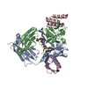
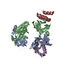
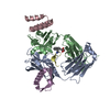
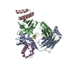
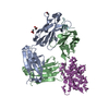
| Title | Trastuzumab Fab v3 in complex with CQFDLSTRRLKC | ||||||
 Components Components |
| ||||||
 Keywords Keywords | IMMUNE SYSTEM / monoclonal antibody / Fab / meditope | ||||||
| Function / homology |  Function and homology information Function and homology informationsymbiont-mediated suppression of host signal transduction pathway via antagonism of host cell surface receptor / IgD immunoglobulin complex / IgA immunoglobulin complex / IgM immunoglobulin complex / IgE immunoglobulin complex / CD22 mediated BCR regulation / Fc epsilon receptor (FCERI) signaling / IgG binding / Classical antibody-mediated complement activation / Initial triggering of complement ...symbiont-mediated suppression of host signal transduction pathway via antagonism of host cell surface receptor / IgD immunoglobulin complex / IgA immunoglobulin complex / IgM immunoglobulin complex / IgE immunoglobulin complex / CD22 mediated BCR regulation / Fc epsilon receptor (FCERI) signaling / IgG binding / Classical antibody-mediated complement activation / Initial triggering of complement / IgG immunoglobulin complex / immunoglobulin mediated immune response / FCGR activation / immunoglobulin binding / Role of LAT2/NTAL/LAB on calcium mobilization / Role of phospholipids in phagocytosis / immunoglobulin complex / Scavenging of heme from plasma / antigen binding / FCERI mediated Ca+2 mobilization / FCGR3A-mediated IL10 synthesis / Regulation of Complement cascade / Antigen activates B Cell Receptor (BCR) leading to generation of second messengers / Cell surface interactions at the vascular wall / B cell receptor signaling pathway / FCGR3A-mediated phagocytosis / FCERI mediated MAPK activation / Regulation of actin dynamics for phagocytic cup formation / Immunoregulatory interactions between a Lymphoid and a non-Lymphoid cell / FCERI mediated NF-kB activation / blood microparticle / Potential therapeutics for SARS / adaptive immune response / immune response / : / extracellular exosome / extracellular region / plasma membrane Similarity search - Function | ||||||
| Biological species |   Homo sapiens (human) Homo sapiens (human) Finegoldia magna (bacteria) Finegoldia magna (bacteria) Staphylococcus aureus (bacteria) Staphylococcus aureus (bacteria)synthetic construct (others) | ||||||
| Method |  X-RAY DIFFRACTION / MOLECULAR REPLACEMENT / Resolution: 2.14 Å X-RAY DIFFRACTION / MOLECULAR REPLACEMENT / Resolution: 2.14 Å | ||||||
 Authors Authors | Bzymek, K.P. / King, J.D. / Williams, J.C. | ||||||
 Citation Citation | Journal: Bioconjug. Chem. / Year: 2018 Title: Template-Catalyzed, Disulfide Conjugation of Monoclonal Antibodies Using a Natural Amino Acid Tag. Authors: King, J.D. / Ma, Y. / Kuo, Y.C. / Bzymek, K.P. / Goodstein, L.H. / Meyer, K. / Moore, R.E. / Crow, D. / Colcher, D.M. / Singh, G. / Horne, D.A. / Williams, J.C. | ||||||
| History |
|
- Structure visualization
| Structure viewer | Molecule: MolmilJmol/JSmol |
|---|
- Downloads & links
-Download
| PDBx/mmCIF format | 6bae.cif.gz | 136.9 KB | Display | PDBx/mmCIF format |
|---|---|---|---|---|
| PDB format | pdb6bae.ent.gz | 103.8 KB | Display | PDB format |
| PDBx/mmJSON format | 6bae.json.gz | Tree view | PDBx/mmJSON format | |
| Others |  Other downloads Other downloads |
-Validation report
| Arichive directory | https://data.pdbj.org/pub/pdb/validation_reports/ba/6baeftp://data.pdbj.org/pub/pdb/validation_reports/ba/6bae | HTTPS FTP |
|---|
-Related structure data
| Related structure data |  6b9yC  6b9zC  6bahC 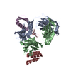 4ioiS C: citing same article ( S: Starting model for refinement |
|---|---|
| Similar structure data |


-Links
 PDBj
PDBj


- Assembly
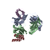
| Deposited unit | 
| ||||||||
|---|---|---|---|---|---|---|---|---|---|
| 1 |
| ||||||||
| Unit cell |
|
-Components
-Antibody , 3 types, 3 molecules ABC
| #1: Antibody | Mass: 23518.004 Da / Num. of mol.: 1 Source method: isolated from a genetically manipulated source Source: (gene. exp.) Homo sapiens (human)Gene: IGKC / Production host:  Cricetulus griseus (Chinese hamster) / References: UniProt: P01834 Cricetulus griseus (Chinese hamster) / References: UniProt: P01834 |
|---|---|
| #2: Antibody | Mass: 23806.670 Da / Num. of mol.: 1 / Mutation: A175C, R217K Source method: isolated from a genetically manipulated source Source: (gene. exp.) Homo sapiens (human)Production host: Cricetulus griseus (Chinese hamster) / References: UniProt: S6B291 |
| #4: Antibody | Mass: 6037.530 Da / Num. of mol.: 1 Source method: isolated from a genetically manipulated source Source: (gene. exp.) Staphylococcus aureus (bacteria) / Gene: spa / Production host: |
-Protein / Protein/peptide / Non-polymers , 3 types, 537 molecules ED
| #3: Protein | Mass: 6907.657 Da / Num. of mol.: 1 / Mutation: T17I, D38A, Y56N, T57H, I58M Source method: isolated from a genetically manipulated source Source: (gene. exp.) Finegoldia magna (bacteria) / Production host: |
|---|---|
| #5: Protein/peptide | Mass: 1496.801 Da / Num. of mol.: 1 / Source method: obtained synthetically / Source: (synth.) synthetic construct (others) |
| #6: Water | ChemComp-HOH / Mass: 18.015 Da / Num. of mol.: 535 / Source method: isolated from a natural source / Formula: H2O |
-Details
| Has protein modification | Y |
|---|
-Experimental details
-Experiment
| Experiment | Method: X-RAY DIFFRACTION / Number of used crystals: 1 |
|---|
- Sample preparation
| Crystal | Density Matthews: 2.66 Å3/Da / Density % sol: 53.82 % |
|---|---|
| Crystal grow | Temperature: 293 K / Method: vapor diffusion, hanging drop / Details: 0.1 M Tris pH 7.5, 24 mM NaCl, 15% PEG 3350 |
-Data collection
| Diffraction | Mean temperature: 100 K |
|---|---|
| Diffraction source | Source: ROTATING ANODE / Type: RIGAKU MICROMAX-007 HF / Wavelength: 1.5418 Å |
| Detector | Type: RIGAKU RAXIS IV++ / Detector: IMAGE PLATE / Date: Dec 7, 2016 |
| Radiation | Protocol: SINGLE WAVELENGTH / Monochromatic (M) / Laue (L): M / Scattering type: x-ray |
| Radiation wavelength | Wavelength: 1.5418 Å / Relative weight: 1 |
| Reflection | Resolution: 2.14→33.53 Å / Num. obs: 37070 / % possible obs: 99.7 % / Redundancy: 5.1 % / CC1/2: 0.995 / Rrim(I) all: 0.152 / Net I/σ(I): 10.3 |
| Reflection shell | Resolution: 2.14→2.2 Å / Redundancy: 5.1 % / Num. unique obs: 2675 / CC1/2: 0.611 / Rrim(I) all: 0.931 / % possible all: 100 |
- Processing
| Software |
| ||||||||||||||||||||||||||||||||||||||||||||||||||||||||||||||||||||||||||||||||||||||||||||||||||
|---|---|---|---|---|---|---|---|---|---|---|---|---|---|---|---|---|---|---|---|---|---|---|---|---|---|---|---|---|---|---|---|---|---|---|---|---|---|---|---|---|---|---|---|---|---|---|---|---|---|---|---|---|---|---|---|---|---|---|---|---|---|---|---|---|---|---|---|---|---|---|---|---|---|---|---|---|---|---|---|---|---|---|---|---|---|---|---|---|---|---|---|---|---|---|---|---|---|---|---|
| Refinement | Method to determine structure: MOLECULAR REPLACEMENT Starting model: 4ioi Resolution: 2.14→33.529 Å / SU ML: 0.22 / Cross valid method: FREE R-VALUE / σ(F): 1.36 / Phase error: 21.18
| ||||||||||||||||||||||||||||||||||||||||||||||||||||||||||||||||||||||||||||||||||||||||||||||||||
| Solvent computation | Shrinkage radii: 0.9 Å / VDW probe radii: 1.11 Å | ||||||||||||||||||||||||||||||||||||||||||||||||||||||||||||||||||||||||||||||||||||||||||||||||||
| Refinement step | Cycle: LAST / Resolution: 2.14→33.529 Å
| ||||||||||||||||||||||||||||||||||||||||||||||||||||||||||||||||||||||||||||||||||||||||||||||||||
| Refine LS restraints |
| ||||||||||||||||||||||||||||||||||||||||||||||||||||||||||||||||||||||||||||||||||||||||||||||||||
| LS refinement shell |
|
